D. Others:
略.
E. Reference:
略.
|
|
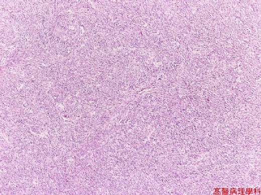
【 Fig. 209-1 (LP)】The tumor is composed of spindle cells in interlacing pattern.
|
|
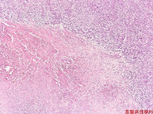
【 Fig. 209-2 (LP)】Focal necrosis is noticed with tumor.
|
|
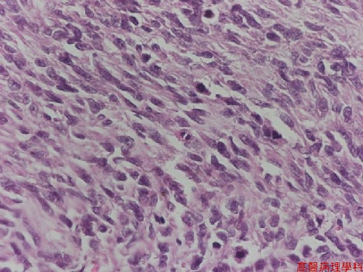
【 Fig. 209-3 (LP)】The tumor cells have pleomorphic nuclei and frequent mitotic figures (>5/50 HPF).
|
|
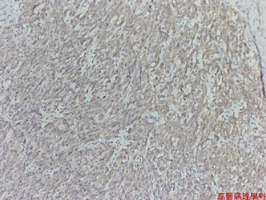
【 Fig. 209-4 (LP)】Immunohistochemical study shows cytoplasmic positive reaction with CD117 (brown color).
![]()
![]()
![]()