《Slide 165.》Meningioma, Meninges
A. Brief Descriptions:
-
Arising from the meningothelial cell of the arachnoid.
-
Histologic pattern: syncytial, fibroblastic, transitional, psammomatous, angiomatous, etc.
B. Gross Findings:
-
Encapsulated,round masses with a well-defined dura base and easily seperated.
-
Firm to fibrous and lack of hemorrhage and necrosis.
-
Hyperostotic reaction in the overlying bone.
C. Micro Findings:
-
Sheets and whorls of polygonal cells, with large, pale spheroid, central nuclei and abundant cytoplasm supported by collagen stroma.
-
Psammoma bodies can also be found.
D. Others:
-
Meningioma起源於subarachnoid的meningothelial cells,所以在腦的表面和腦室內,甚至沿著spinal cord都可以發生meningioma。常見於parasagittal aspect of the convexity,dura over the lateral convexity,the wing of the sphenoid,olfactory groove,sella turcica,foramen magnum.
-
Slow growing and expressing progesterone receptors.
-
在染色體第22對上的long arm缺失。
-
特殊染色: EMA positive and CEA positive.
E. Reference:
-
Robbins Pathologic Basis of Disease, 6th ed. P.1350-1351.
|
|
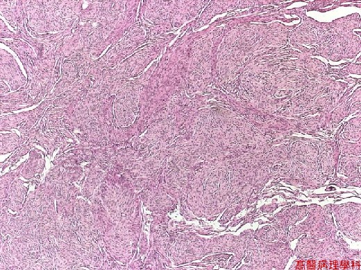
【 Fig. 165-1 (LP)】The tumor in section is whorled and lobulated.
|
|
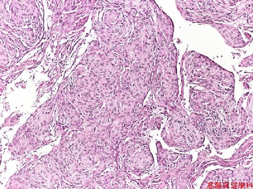
【 Fig. 165-2 (LP)】
|
|
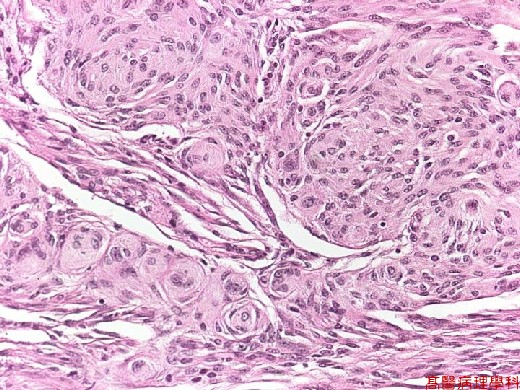
【 Fig. 165-3 (LP)】Meningotheliomatous cells with polygonal to spindle shape and whorl appearance, and sycytoplasmic cells in the center of whorl.
|
|
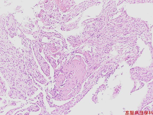
【 Fig. 165-4 (LP)】Mineralized psammoma body.
![]()
![]()
![]()