《Slide
152.》Chondrosarcoma,
dedifferentiated,
Rib
A. Brief Descriptions:
-
A biphasic tumor consisting of a poorly differentiated sarcomatous component juxtaposed to an otherwise typical low-grade chondrosarcoma.
-
Peak age: sixth decade with most common lesions in femur and pelvis.
-
Radiographic features: an aggressive area superimposed on typical chondrosarcoma.
B. Gross Findings:
-
Lobulated, gray-white hyaline cartilage coexists with a well circumscribed soft tumor mass.
C. Micro Findings:
-
Lobulated, hyaline cartilaginous lesion.
-
Cellular lesion (chondrocytes) with pleomorphic, hyperchromatic or binucleated nuclei cells.
D. Others:
略.
E. Reference:
-
Robbins Pathologic Basis of Disease, 6th ed. P1240-1242.
|
|
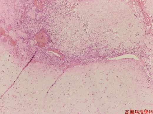
【 Fig. 152-1 (LP)】 Hyaline cartilaginous lesion (lower) surrounded by spindle cells (right upper).
|
|
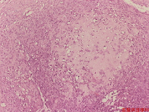
【 Fig. 152-2
(LP)】 Chondroid lesions surrounded by small blue cells with hyperchromatic
nuclei.
|
|
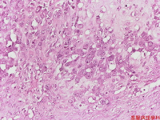
【 Fig. 152-3 (LP)】 Crowed chondrocytes with pleomorphic, hyperchromatic or binucleated nuclei pattern.
|
|
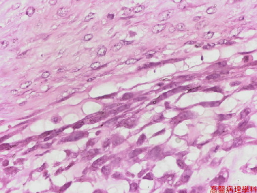
【 Fig. 152-4 (HP)】(Upper) chondrocytes with pleomorphic, hyperchromatic or binucleated nuclei pattern. (Lower) Spindle cell stroma showed marked cellular atypia with atypical mitotic figures.
![]()
![]()
![]()