《Slide 3.》Necrosis of cerebellum, Cerebellum
A. Brief Descriptions:
-
Encephalomalacia: necrosis of the brain following by liquefaction & secondary cavitation.
-
Stages:
-
Cortical pallor with ischemic change in neurons.
-
Softening with granular foamy cells (Gitter cells).
-
Cystic formation & glial scarring.
-
C. Micro Findings:
-
Congestion, hemorrhage, or hemosiderin deposits.
-
Destruction of structure with heavy infiltration by neutrophils, lymphocytes....
-
Necrosis & granular foamy cells ( from phagocytic microglial cells, Gitter cells ).
-
Gitter cells: vacuolated, eosinphilic or granular cytoplasm with eccentrically located nuclei.
D. Reference:
-
Robbins Pathologic Basis of Disease, 6th ed. P.15-18.
|
|
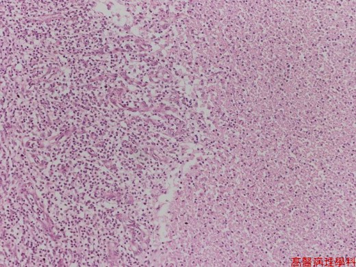
【 Fig. 3-1 (LP)】The necrotic area (right half) is surrounded by granulation tissue in the left.
|
|
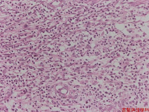
【 Fig. 3-2 (LP)】The granulation tissue is composed of regenerated vasculature, fibroblasts and inflammatory cells.
|
|
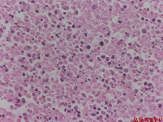
【 Fig. 3-3 (HP)】The necrotic area is composed of cell debris (pinkish color) and nuclear dusts (dark purple or blue color).
![]()
![]()
![]()