B. Gross Findings:
-
Blackish discoloration of gastric mucosa, hemorrhage , ulceration, or perforation.
C. Micro Findings:
-
Hemorrhage, congestion, coagulative necrosis of mucosa with hemosiderin or carbon deposit.
-
Necrotizing necrosis with inflammatory infiltration.
-
Variant pictures due to agents, amounts & concentration (depth of layers involved).
D. Others:
-
Causes :ingestion of corrosive agents (lye, acids) with dramatic injury to the esophagus and stomach.
E. Reference:
-
Robbins Pathologic Basis of Disease, 6th ed. P.789-790.
|
|
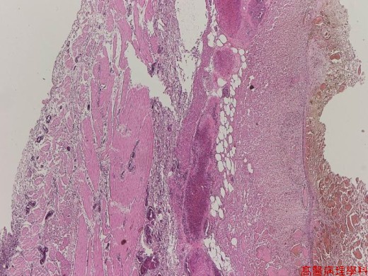
【 Fig. 6-1 (LP)】Coagulative necrosis of mucosa and submucosa with relatively preserved muscularis propria.
|
|
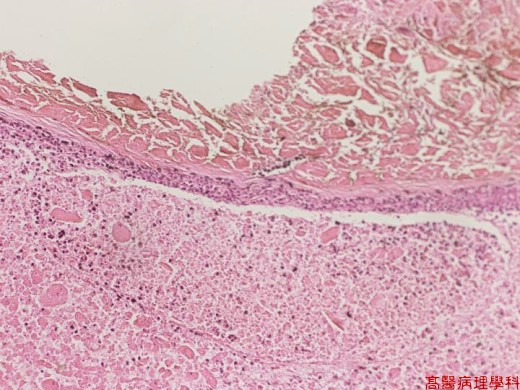
【 Fig. 6-2 (LP)】Coagulative necrosis of mucosa (upper) and submucosa (lower). Note the relatively preserved contour of mucosal glands with loss of individual nuclei.
|
|
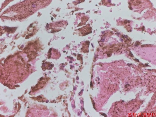
【 Fig. 6-3 (LP)】Coagulative necrosi of mucosal glands with relatively preserved glandular contour but without nucleus. A residual gland is seen in the center.
|
|
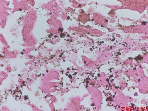
【 Fig. 6-4 (LP)】There are many brown to black variable sized particles deposit in coagulative necrotic area.
|
|
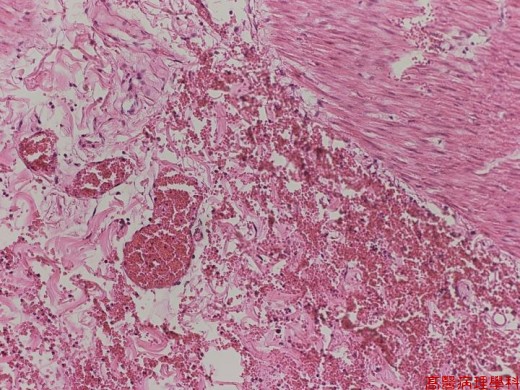
【 Fig. 6-5 (LP)】Congestion and hemorrhage in muscularis propria.
|
|
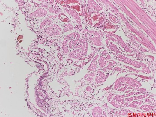
【 Fig. 6-6 (LP)】The serosal surface (left) is coated with fibrinoid substance with inflammatory cells infiltrate. Note diffuse congestion of vessels.
![]()
![]()
![]()