《Slide 203.》Tuberculosis, Lung
A. Brief Descriptions:
-
Caused by Mycobacterium tuberculosis.
B. Gross Findings:
-
Primary TB – Ghon complex.
-
Secondary TB – in the apex as a small focus of consolidation.
C. Micro Findings:
-
Granulomas (tubercles):
-
Central caseation necrosis.
-
Epithelioid histiocytes.
-
Multinucleated Langhans’ giant cells.
-
Fibroblasts and lymphocytes.
-
E. Reference:
-
Robbins Pathologic Basis of Disease, 6th ed. P.379-352.
|
|
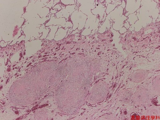
【 Fig. 203-1 (LP)】Multiple confluent or separated nodules are seen in lung parenchyma. Note thickened and congested interstitium. The alveoli show emphysematous change.
|
|
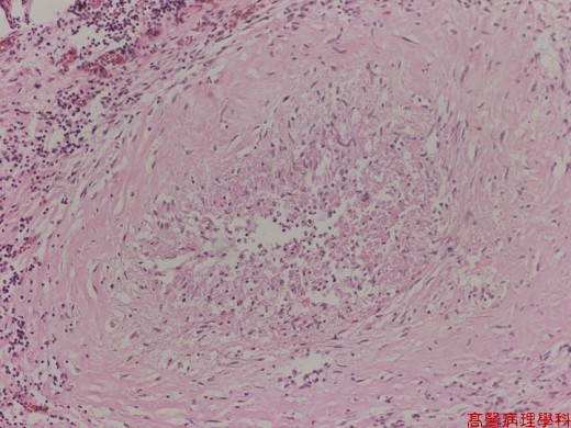
【 Fig. 203-2 (LP)】Caseating granuloma (tubercle). The center of the tubercle contains caseous necrotic debris (amorphous eosinophilic substance) and is surrounded by epithelioid histiocytes and lymphocytes.
|
|
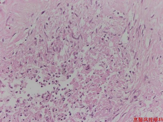
【 Fig. 203-3 (LP)】The center of the tubercle is composed of caseating necrotic debris. Note the elongated epithelioid histiocytes around the caseating center.
|
|
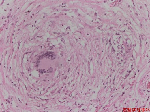
【 Fig. 203-4 (LP)】A caseous granuloma with caseating necrotic center, epithelioid histiocytes and a typical Langhans’ giant cell (middle left). Note the nuclei of the Langhans’ type giant cell lined in horseshoe arrangement.
|
|
.jpg)
【 Fig. 203-5 (HP, acid fast stain)】The acid fast stain highlights bacilli of Mycobacterium species (pink color) (Most commonly M. tuberculosis).
![]()
![]()
![]()