B. Gross Findings:
-
All infarction tend to forming wedge shaped occluded vessel at apex and the periphery of the organ forming the base.
-
Spongy and cyanotic in red infarct at first.
-
The margin of both type become progressively better defined in the course of few days.
C. Micro Findings:
-
Irregular fibrous bundles replaced the cardiac muscle as chronic myocardial infarction.
-
Interrupted and separated cardiac muscle by fibrous tissue.
-
Proliferation of capillary in the interstitium.
D. Others:
-
The coagulative necrosis may not detectable for the first 4~8 hrs.
-
Sequential changes in myocardial infarction:
|
time |
gross |
light microscopy |
|
1 ~ 4 hrs |
none |
none |
|
4 ~ 12 hrs |
none |
begin coagulative necrosis, neutrophil infiltrates ; edema, hemorrhage. |
|
18 ~ 24 hrs |
pallor |
continuing coagulation necrosis marginal contraction band necrosis |
|
24 ~ 72 hrs |
pallor, sometimes hyperemia |
loss of nuclei and striation heavy infiltration of neutrophils |
|
3 ~ 7 days |
hyperemic border central yellow-brown |
disintegration of dead myofibers macrophages present onset of marginal fibrovascular response |
|
10 days |
red-brown & depressed yellow & soft vascularized margins |
well-develop necrotic changes prominent fibrovascular reaction in margin |
|
7th weeks |
scarring |
fibrotic |
E. Reference:
-
Robbins Pathologic Basis of Disease, 6th ed. P.71.
|
|
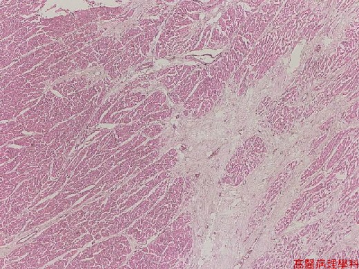
【 Fig. 23-1 (LP)】Irregular fibrous bundles (left) replaced the cardiac muscles.Granulation tissue (right) with a rich vascular network and early collagen deposition.
|
|
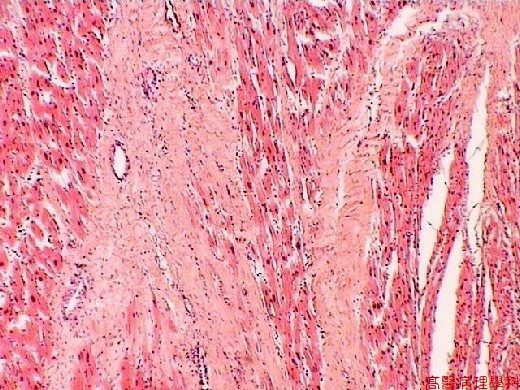
【 Fig. 23-2 (HP)】Fibrous bundles arrange in a parallel fashion with capillaries and minimal inflammation; note separated and interrupted cardiac muscles.
|
|
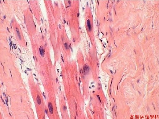
【 Fig. 23-3 (HP)】Fibrous bundles arrange in a parallel fashion with capillaries and minimal inflammation; note separated and interrupted cardiac muscles.
![]()
![]()
![]()