B. Gross Findings:
略.
C. Micro Findings:
-
Heart failure cell presented in alveolar spaces.
-
Congestion and hemorrhage.
-
Anthracotic deposits in alveolar septi.
D. Others:
略.
E. Reference:
-
Robbins Pathologic Basis of Disease, 6th ed. P.549.
|
|
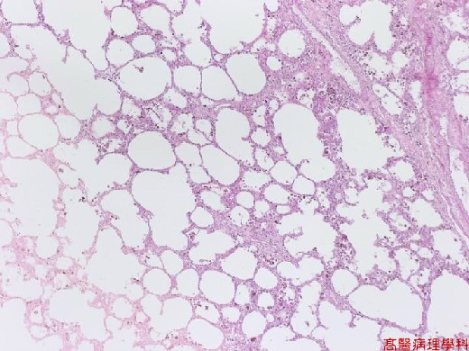
【 Fig. 26-1 (LP)】The alveolar spaces are filled with exudate and brownish heart failure cells; note entrapped air bubbles and thickened alveolar septi.
|
|
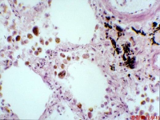
【 Fig. 26-2 (HP)】Heart failure cells as hemosiderin laden macrophages in the alveolar spaces; note anthracosis (dark brown carbon dusts) of the interstitium (right upper).
![]()
![]()
![]()