《Slide 25.》Splenic infarction, Spleen
A. Brief Descriptions:
-
Nearly 99% of infarcts are caused by thromboembolic events, and almost all are the result of arterial occlusions.
-
White infarcts are encountered with arterial occlusion and in solid tissues.
B. Gross Findings:
-
Recent infarcts are hemorrhagic,whereas older, more fibrotic infarcts are pale yellow-gray.
C. Micro Findings:
-
Necrotic area with homogenous pinkish appearance.
-
Hematoidin crystals can be found in this section.
-
Inflammatory cells seated on the margin of infarct area.
D. Others:
略.
E. Reference:
-
Robbins Pathologic Basis of Disease, 6th ed. P.689-690.
|
|
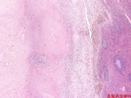
【 Fig. 25-1 (LP)】Splenic infarction (left) with homogenous pinkish appearance.
|
|
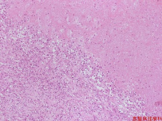
【 Fig. 25-2 (LP)】Inflammatory cells seated on the margin of infarct area.
|
|
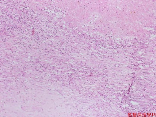
【 Fig. 25-3 (LP)】Inflammatory cells.
|
|
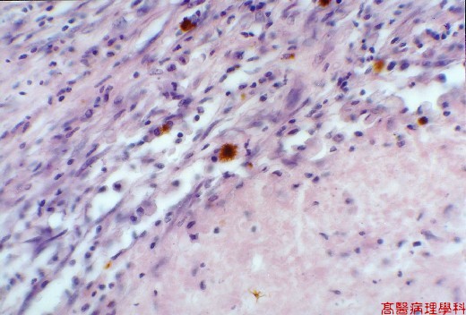
【 Fig. 25-4 (HP)】Hemosiderin laden macrophages in the more fibrous areas (junction of infarction ).
![]()
![]()
![]()