C. Micro Findings:
-
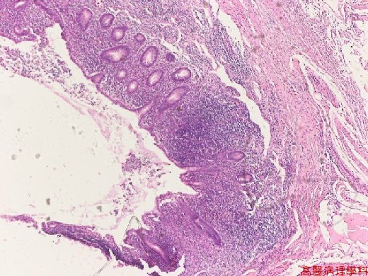
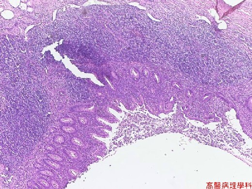
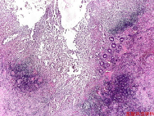
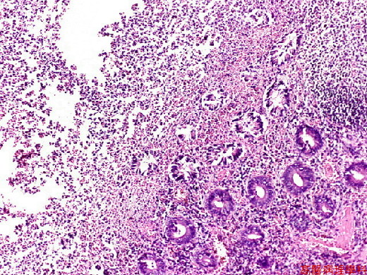
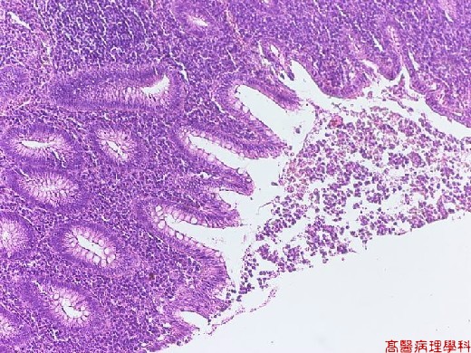
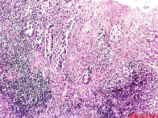
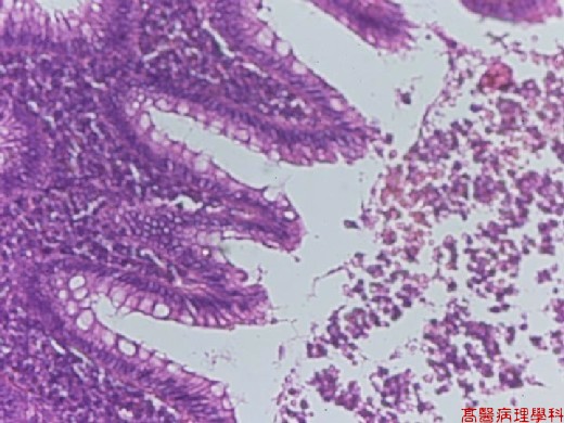
Mucosal ulceration & infiltration by PMNs, eosinophils, plasma cells, &lymphocytes throughout all layers & frequently into serosa.
-
More advanced stage, the inflammatory process involved the full thickness of wall,with partial necrosis or infarction of wall (perforated areas).
D. Others:
-
Classified into acute, suppurative, & gangrenous stages.
site
acute
suppurative
gangrenous
mucosa
neutrophils
suppurative necrosis
hemorrhagic ulceration
wall
neutrophils
suppurative necrosis
green-black necrosis
serosa
congested blood vessels
fibrinous exudates
purulent exudates
green-black necrosis
E. Reference:
-
Robbins Pathologic Basis of Disease, 6th ed. P.839-840.
|
|
【 Fig. 17-1 (LP)】
|
|
【 Fig. 17-2 (LP)】
|
|
【 Fig. 17-3 (LP)】
|
|
【 Fig. 17-4 (LP)】
|
|
【 Fig. 17-5 (LP)】
|
|
【 Fig. 17-6 (LP)】
|
|
【 Fig. 17-7 (HP)】
