E. Reference:
Robbins Pathologic Basis of Disease, 6th ed. P.783-786.
|
|
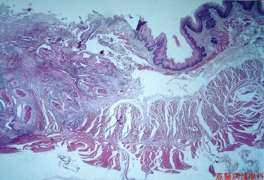
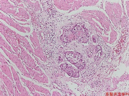
【 Fig. 177-1 (2X)】Squamoid cancer cells invaded into muscular layer seen in this low power field.
|
|
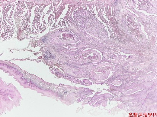
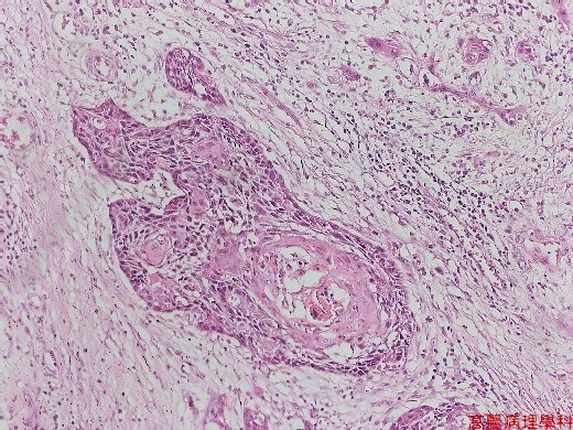
【 Fig. 177-2 (4X)】Squamoid cancer cells grew from surface and invaded into muscular layer seen in this view.
|
|
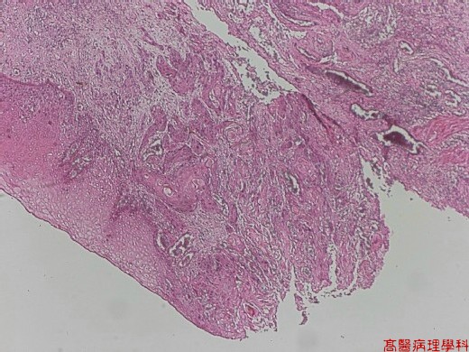
【 Fig. 177-3 (4X)】Ulcerated squmaoid cancer cells grew into submuosa.
|
|
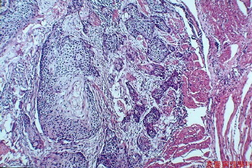
【 Fig. 177-4 (10X)】Squamoid cancer cells invaded into muscular layer.
|
|
【 Fig. 177-5 (10X)】Squamoid cancer cells with keratinizing.
|
|
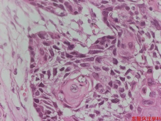
【 Fig. 177-6 (20X)】Squamoid cancer cells with keratinizing.
|
|
【 Fig. 177-7 (40X)】Intercellular bridge seen in high power view.
![]()
![]()
![]()