E. Reference:
Robbins Pathologic Basis of Disease, 6th ed. P.1021-1022.
|
|
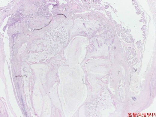
【 Fig. 39-1 (1X)】Cystic & multiloculated lesion.
|
|
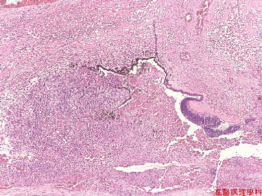
【 Fig. 39-2 (4X)】Epithelial component seeen.
|
|
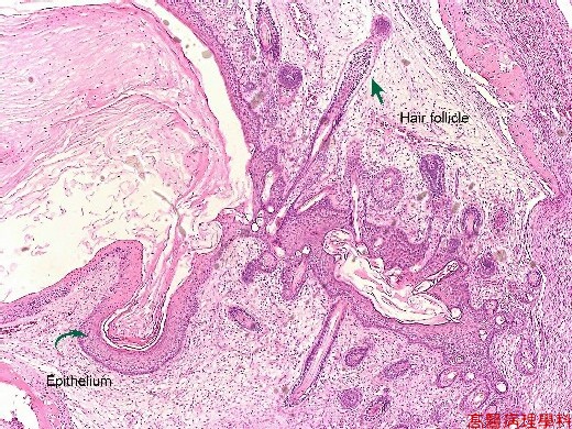
【 Fig. 39-3 (4X)】Skin component seen in this view.
|
|
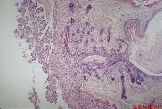
【 Fig. 39-4 (4X)】Choroid plexus-like tissue seen in left view.
|
|
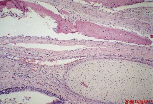
【 Fig. 39-5 (10X)】Catilage and bone seen in this view.
|
|
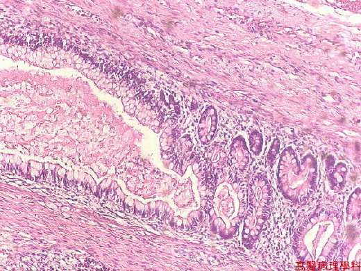
【 Fig. 39-6 (10X)】Mature goblet cells seen in this picture .
![]()
![]()
![]()