《Slide 27.》 Squamous cell carcinoma, Skin
A. Brief Descriptions:
-
Most often occurs on sun-exposed skin.
-
Risk factors: sunlight, industrial carcinogen(tars and oils),chronic ulcers and draining osteomyelitis ,old burn scars, ingestion of arsenicals, ionizing radiation, and (in the oral cavity) tobacco and betel nut chewing.
B. Gross Findings:
A shallow ulcer surrounded by a wide, elevated indurated border, occasionally raised, fungoid, verrucous lesions without ulceration.
C. Micro Findings:
-
Irregular masses of epithelial cells proliferate downward into the dermis.
-
Individual tumor masses composed in varying proportion of normal or atypical squamous cells.
-
Atypia: varying size & shape, hyperplasia, hyperchromatism, absence of intercellular bridge, presence of atypical mitosis & dyskeratotic cells.
-
Dyskeratotic cells: individual cell keratinization with large, round & deep eosinophilic cytoplasm & hyperchromatic nuclei.
-
Well differentiated cells with intercellular bridge & keratin pearl formation & sheets of partially keratinized cells.
-
Keratin pearl: a rounded nest of squamous epithelium in which the cells are arranged in concentric circles surrounding a central focus of acellular keratin.
D. Others:
略
E. Reference:
Robbins Pathologic Basis of Disease, 6th ed. P.1184-1186.
|
|
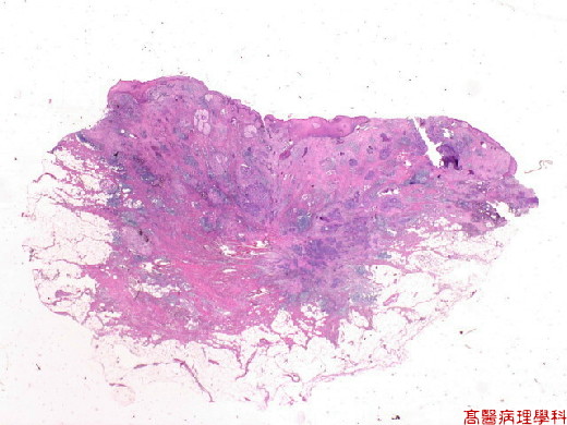
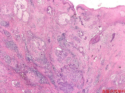
【 Fig. 27-1 (1X)】.Cancer cells infiltrate from surface down into subcutis in the low power field.
|
|
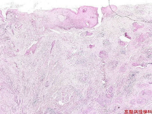
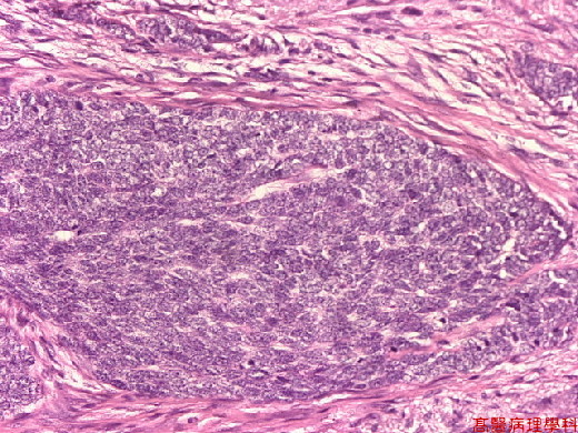
【 Fig. 27-2 (4X)】.Cancer nests are seen in dermis.
|
|
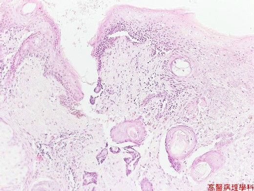
【 Fig. 27-3 (2X)】Cancer nests are seen in dermis.
|
|
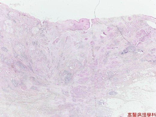
【 Fig. 27-4 (4X)】Cancer nests are seen in subcutis.
|
|
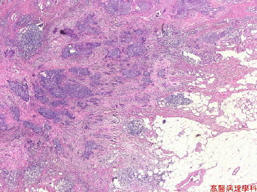
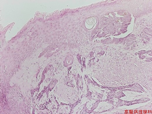
【 Fig. 27-5 (4X)】Cancer cells with tendency to keratinization.
|
|
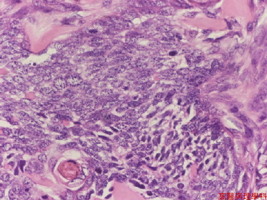
【 Fig. 27-6 (10X)】Keratinizing cancer nests seen in superficial dermis.
|
|
【 Fig. 27-7 (20X)】Squamoid cancer cells with keratinization and tendency to keratin pearl formation.
|
|
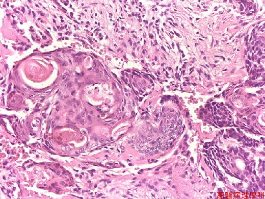
【 Fig. 27-8 (20X)】Keratin pearl formation is seen above.
|
|
【 Fig. 27-9 (20X)】Nest of hyperchromatic cancer cells with high nuclear and cytoplasmic ratio.
|
|
【 Fig. 27-10 (20X)】Keratin pearl formation is seen in left lower field.
|
|
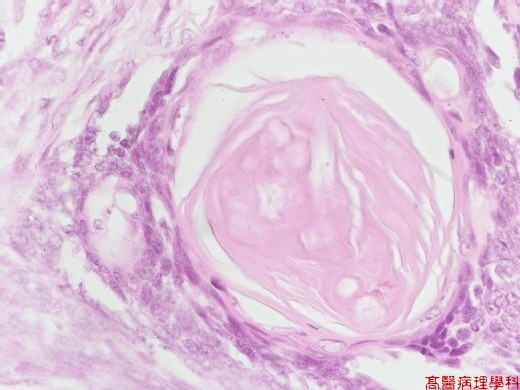
【 Fig. 27-11 (40X)】Keratin pearl seen in this high power field.
![]()
![]()
![]()