Slide 34c.Lipoma, Colon

A. Brief Descriptions

  1. Rare in intestinal tumor.

  2. More commonly seen in large intestine than in small intestine.

B. Gross Findings

  1. Lipoma of small bowel: submucosal, bright yellow, round, encapsulated, may bulge upward into mucosal surface.

  2. Lipoma of soft tissue: bright yellow, round, encapsulated fat, but maybe a infiltrated pattern in deeper structure.

  3. Combined with other components.

     Ex: angiolipoma, fibrolipoma, angiomyolipoma, myxolipoma.

  4. Areas of infarction, necrosis, calcification, osseous, or cartilaginous metaplasia may be found. (Not in our slide).

C. Micro Findings

     Mature adipose tissue (univacular mature adipocytes) with no cellular atypia.

D. Others:

   略.

E. Reference

     Robbins Pathologic Basis of Disease, 6th ed.  P.838-839.
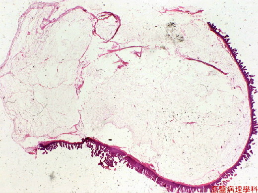


Fig. 34-1 (1X)Well circumscribed tumor in submuosal layer.
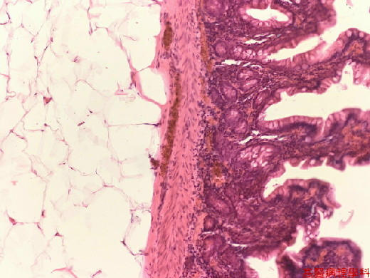


Fig. 34-2 (10X)Submucosal, bright yellow, round, encapsulated tumor.
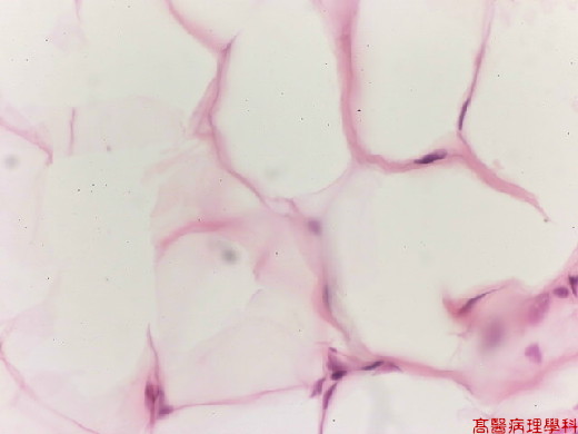


Fig. 34-3 (40X).Mature adipocytes seen in this picture.