¡mSlide 70.¡nTransitional cell carcinoma, Ureter
A. Brief Descriptions¡G
-
Male predominance.
-
Most common in order individuals and tobacco and industrial carcinogen exposure are risk factors.
-
Symptoms and signs including hematuria and flank pain.
B. Gross Findings¡G
Varied from purely papillary to nodular, or flat pattern, even mixed architecture.
C. Micro Findings¡G
Papillary & non-papillary type
-
In papillary type (exophytic lesion): a thin core of fibrovascular tissue covered by layers of tumor cells, like a molded cauliflower.
-
In non-papillary type: a smooth or slightly bulging lesion with extensive invasion.
D. Others:
According to WHO grading system:
-
Grade 1: increase in number of layers of cells showing some atypia.
-
Grade 2: The number of layers of cells increased, as in the number of mitosis. Cancer cells show pleomorphism, hyperchromatism, and loss of polarity but are still recognizable as of transitional origin.
-
Grade 3: Many of the tumor cells show anaplastic change with loosening and fragmentation of the superficial layers of the cells. Giant cancer cells may be present. Focal squamous or glandular metaplasia.
E. Reference¡G
Robbins Pathologic Basis of Disease, 6th ed. P.999.
¡@
|
|
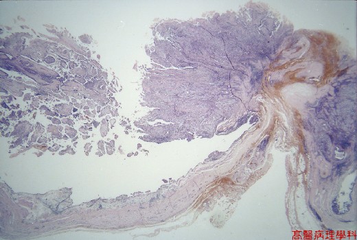
¡i Fig. 70-1 (1X)¡jPapillary tumor grow seen in this low power field.
¡@
|
|
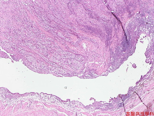
¡i Fig. 70-2 (2X)¡jTumor seen in upper field.
¡@
|
|
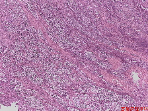
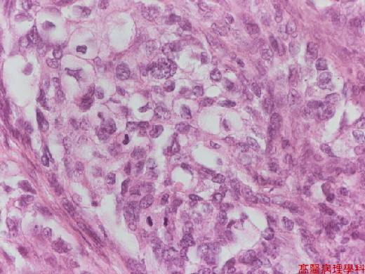
¡i Fig. 70-3 (4X)¡jSolid tumor cells with clear cytoplasm.
¡@
|
|
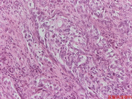
¡i Fig. 70-4 (20X)¡jSolid tumor cells with clear cytoplasm.
¡@
¡@
|
|
¡i Fig. 70-5 (40X)¡jHyperchromatic cancer cells with clear cytoplasm and mitoses can be seen in lower picture view.
