¡mSlide 71.¡nSeminoma (classic type), Testis
A. Brief Descriptions¡G
The most common germ cell tumors.
B. Gross Findings¡G
Enlarged testis with a well-defined homogenous pale pinkish/white to light yellow tumor may contain
shapely circumscribed zones of necrosis.
C. Micro Findings¡G
-
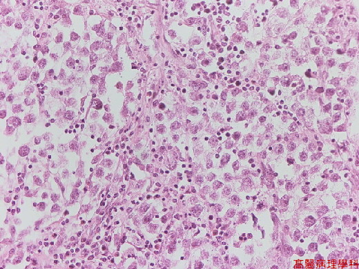
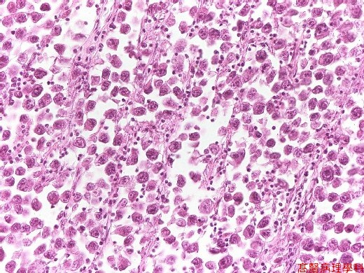
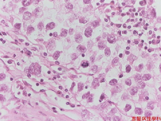
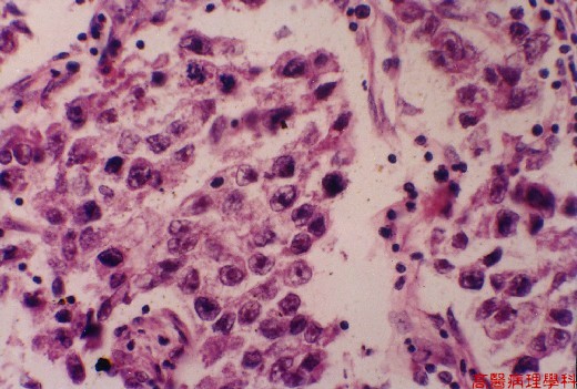
Uniform spheroidal cells with abundant clear cytoplasm, shapely outlined cell membrane, a large centrally located nucleus & clumped chromatin pattern.
-
Prominent nucleolus with amphophilic staining, apparent multiplicity, elongated shaped & irregular contours.
-
Mitosis is variable.
-
Tumor cells arranged in nests outlined by fibrous bands which are infiltrated by lymphocytes & plasma cells.
D. Others:
The most common single type of primary testicular tumor, in two forms: classic type (93%) &
spermatocytic variety.
E. Reference¡G
Robbins Pathologic Basis of Disease, 6th ed. P.1019-1020.
¡@
|
|
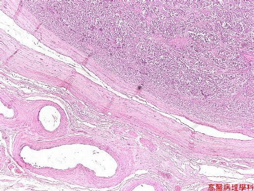
¡i Fig. 71-1 (1X)¡jTumor cells seen in upper field.
¡@
|
|
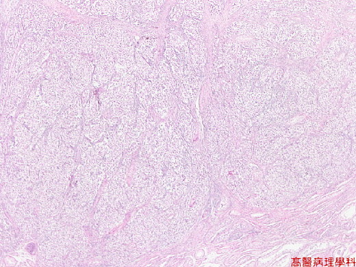
¡i Fig. 71-2 (2X)¡jUniform spheroidal cells with abundant clear cytoplasm, shapely outlined cell membrane.
¡@
|
|
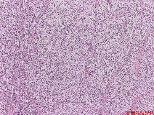
¡i Fig. 71-3 (3X)¡jUniform spheroidal cells with abundant clear cytoplasm, shapely outlined cell membrane.
¡@
|
|
¡i Fig. 71-4 (10X)¡jTumor cells arranged in nests outlined by fibrous bands which are infiltrated by lymphocytes & plasma cells.
¡@
¡@
¡@
|
|
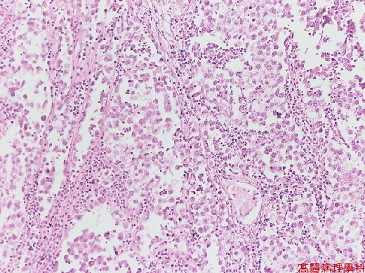
¡i Fig. 71-5 (20X)¡jTumor cells outlined by fibrous bands which are infiltrated by lymphocytes & plasma cells.
¡@
|
|
¡i Fig. 71-6 (20X)¡jTumor cells outlined by fibrous bands which are infiltrated by lymphocytes & plasma cells.
¡@
|
|
¡i Fig. 71-7 (40X)¡jUniform spheroidal cells with abundant clear cytoplasm.
¡@
|
|
¡i Fig. 71-8 (40X)¡jUniform spheroidal cells with abundant clear cytoplasm.
¡@
¡@
