《Slide 75.》Condyloma acuminatum, Penis
A. Brief Descriptions:
-
Benign tumor caused by several types of human papillomavrus (HPV).
-
It is related to the common wart ( verruca vulgaris ) and may occur on any moist muco-cutaneous surface of the external genitals in either sex.
B. Gross Findings:
Single or multiple sessile or pedunculated, red papillary excrescences that vary from 1 to several millimeters in diameter, occasionally coalesce into cauliflowerlike masses.
C. Micro Findings:
-
A branching , villous or papillary connective tissue stroma covered by a thickened hyperplastic epithelium.
-
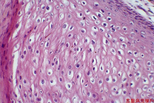
The epithelium shows considerable superficial hyperkeratosis, parakeratosis and thickening of the underlying epidermis (acanthosis) with thickening and elongation of the rete ridges.
-
Distinct perinuclear clear vacuolization of the prickle cells (koilocytosis) as characteristic of HPV infection.
-
These vacuolated epithelial cells are relatively large and possess a hyperchromatic, round nucleus.
-
The basement membrane is intact without invasion of the underlying stroma.
-
The stroma (dermis) appears edematous with dilated capillaries and a moderately dense, chronic inflammatory infiltrate.
D. Others:
略.
E. Reference:
Robbins Pathologic Basis of Disease, 6th ed. P.1012-1013.
|
|
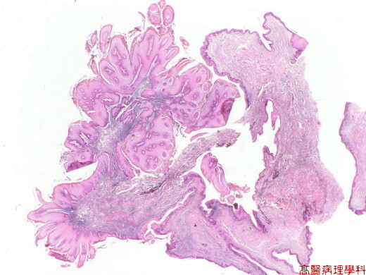
【 Fig. 75-1 (1X)】A branching , villous or papillary connective tissue stroma covered by a thickened hyperplastic epithelium.
|
|
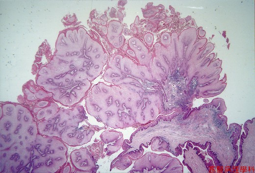
【 Fig. 75-2 (2X)】A branching , villous or papillary connective tissue stroma covered by a thickened hyperplastic epithelium.
|
|
【 Fig. 75-3 (4X)】Distinct perinuclear clear vacuolization of the prickle cells (koilocytosis) as characteristic of HPV infection..
|
|
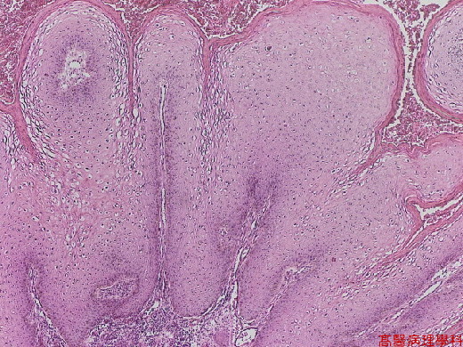
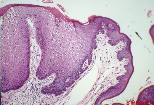
【 Fig. 75-4 (10X)】The epithelium shows considerable superficial hyperkeratosis, parakeratosis and thickening of the underlying epidermis (acanthosis) with thickening and elongation of the rete ridges.
|
|
【 Fig. 75-5 (20X)】Koilocytic atypia seen in this view.
|
|
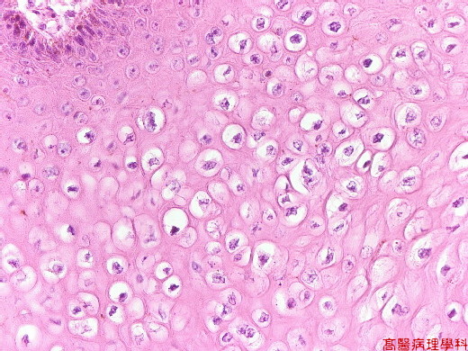
【 Fig. 75-6 (40X)】Koilocytic atypia seen in this view.
![]()
![]()
![]()