《Slide 74.》Yolk sac tumor (endodermal sinus tumor), Testis
A. Brief Descriptions:
-
A neoplasm composed of pure yolk sac elements ,is the most common tumor of infancy.
-
Yolk sac tumors of infancy have a good prognosis.
B. Gross Findings:
A soft consistency and a microcystic appearance on cross section.
C. Micro Findings:
-
Intermingling of epithelial and mesenchymal elements in a characteristic organoid fashion.
-
Microcystic, glandular-alveolar, and papillary formation.
-
Many cystic spaces lined by a very flattened, endothelial-like layer of cells.
-
The stroma can be quite cellular, spindle shaped cells or myxoid.
-
Perivascular Schiller-Duval bodies are the most distinctive feature:
-
papillary projections covered by two distinct cell layers separated from each other by a delicate fluorovascular stroma.
-
-
Hyaline intracytoplasmic or extracytoplasmic round inclusions.
-
eosinophilic, PAS stain positive and diastase resistance.
-
immunocytochemistry forá -fetoprotein.
-
D. Others:
-
Developed teratoma mimicking embryonal yolk sac tissue.
-
Elevated serum α -fetoprotein (tumor marker).
-
Two distinct circumstances:
-
A pure form - with the classic organoid appearance (described by Teilum) in infants and children, with an excellent prognosis.
-
A component of a mixed germ cell tumor in adults, with a lesser differentiated and more malignant appearance, and worse prognosis.
-
E. Reference:
Robbins Pathologic Basis of Disease, 6th ed. P.1075-1076.
|
|
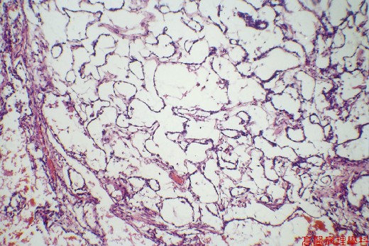
【 Fig. 74-1 (2X)】Tumor cells in cystic pattern.
|
|
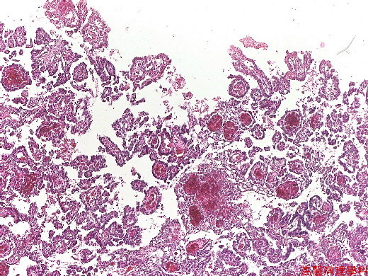
【 Fig. 74-2 (2X)】Papillary pattern with Schiller-Duval bodies.
|
|
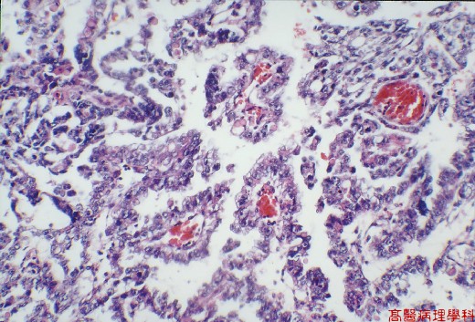
【 Fig. 74-3 (4X)】Perivascular Schiller-Duval bodies are the most distinctive feature.
|
|
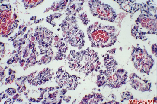
【 Fig. 74-4 (4X)】Perivascular Schiller-Duval bodies are the most distinctive feature.Intracytoplasmic and extracytoplasmic inclusion bodies are seen.
|
|
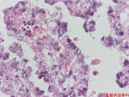
【 Fig. 74-5 (10X)】Intracytoplasmic and extracytoplasmic inclusion bodies are seen.
|
|
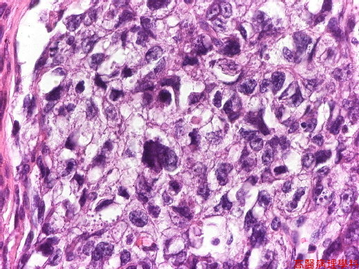
【 Fig. 74-6 (40X)】Hyperchromatic cancer cells in solid pattern.
![]()
![]()
![]()