《Slide 99.》Hodgkin lymphoma , Lymph node
A. Brief Descriptions:
-
Hokgdin lymphoma is characterized morphologically by the presence of distinctive neoplastic giant cells, RS cells, which induce the accumulation of reactive lymphocytes, histiocytes, and granulocytes.
-
Classification:
-
Nodular sclerosis
-
Mixed cellularity
-
Lymphocyte predominance
-
Lymphocyte depletion
-
B. Gross Findings:
-
Enlarged and encapsulated lymph noeds with fish flesh cut surfaces.
C. Micro Findings:
-
Diffuse effacement of lymph nodes by a heterogeneous cellular infiltrate.
-
Moderate amount of RS cells and Hodgkin cells.
-
Lacunar cells, mummified cells.
-
Background: benign-looking small lymphocytes, histiocytes.
-
Mild interstitial fibrosis and few bands of collagen (especially along vessels).
D. Others:
-
Reed-Sternberg cell (RS cell)
-
Classic RS cell
-
Large (15~45m), binucleate or bilobed, with the two halves often appearing as “mirror-image” of each other.
-
Nucleus: may be multiple or multilobate, contains large, inclusion-like, owl-eyed nucleoli, generally surrounded by a clear halo, that are about the size of a small lymphocyte (5~7 m).
-
Cytoplasm: abundant amphophilic.
-
-
Variants of RS cells (1)
-
Mononuclear variants
-
Hodgkin cells.
-
A single round or oblong nucleus with a large inclusion-like nucleolus.
-
-
Lacunar variants
-
Folded or multilobate nuclei surrounded by abundant pale cytoplasm which distrupted during the cutting of sections , leaving the nucleus sitting in an empty hole.
-
Predominant in nodular sclerosis type.
-
-
Lymphocytic and histiocytic variants (L+H cells)
-
Polypoid nuclei resembling popcorn kernels, inconspicuous nucleoli, and moderately abundant cytoplasm.
-
Specific to the lymphocyte predominance subtype.
-
-
Mummified cells
-
Degenerate L+H cells or RS cells.
-
Large cell with darkly staining eosinophilic cytoplasm and a dense pyknotic nucleus.
-
-
-
E. Reference:
-
Robbins Pathologic Basis of Disease, 6th ed. P.670-675.
|
|
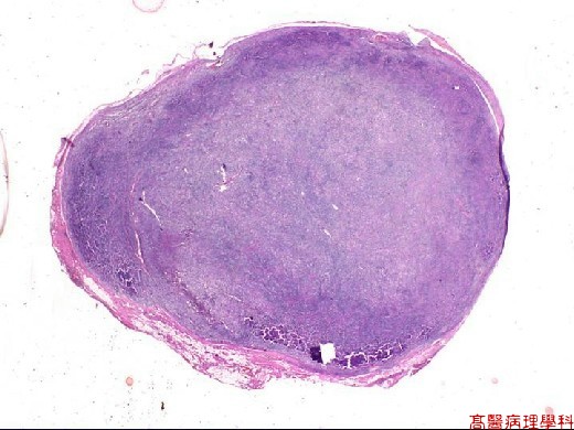
【 Fig. 99-1 (LP)】Diffuse effacement of lymph node by neoplastic lymphoid infiltrate.
|
|
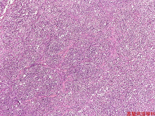
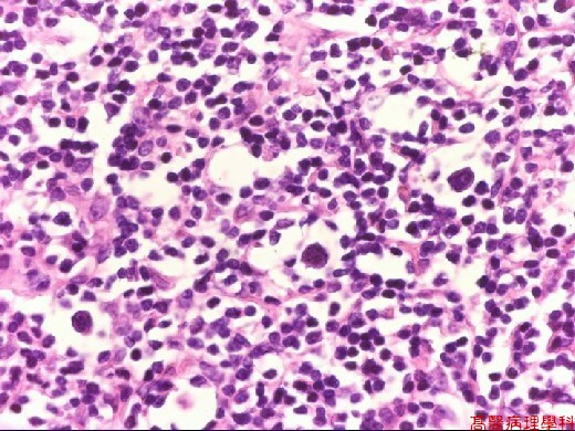
【 Fig. 99-2 (LP)】Heterogeneous cellular infiltrate.
|
|
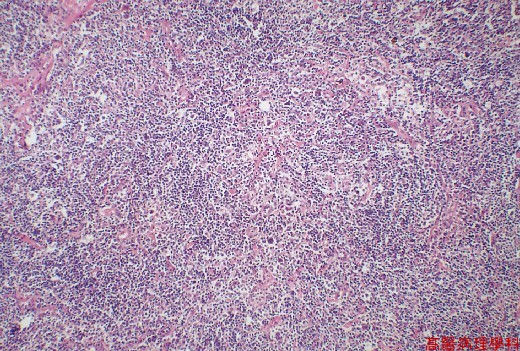
【 Fig. 99-3 (LP)】Neoplastic Hodgkin lymphoma in the background of lymphocytes and histiocytes.
|
|
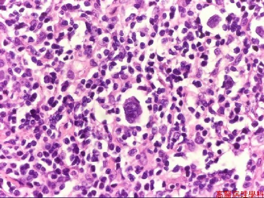
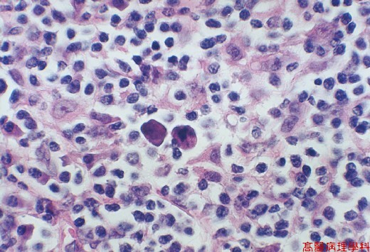
【 Fig. 99-4 (HP)】Classical Reed-Sternberg cells with large "mirror-image" like nuclei and eosinophilic owl-eye nucleoli surrounded by a clear halo.
|
|
【 Fig. 99-5 (HP)】Lacunar cells with delicate folded nuclei surrounded by abundant pale cytoplasm.
|
|
【 Fig. 99-6 (HP)】Mummified cells with darkly staining eosinophilic cytoplasm and a dense pyknotic nucleus.
![]()
![]()
![]()