《Slide 104.》Langerhans cell histiocytosis, Soft tissue
A. Brief Descriptions:
-
Definition: Langernans cell histiocytosis is a neoplastic proliferaion of Langerhans cells, with expresion of CD1a, S-100-protein, and the presence of Birbeck granules by ultrastructural examination.
-
Synonyms: Histiocytosis X, Langerhans cell granulomatosis.
-
Clinical variants: Letterer-Siwe disease, Hand-Schuller-Christian disease, and solitary eosinophilic granuloma.
B. Gross Findings:
略.
C. Micro Findings:
-
Langerhans cells
-
10~15 m, grooved, folded, indented, or lobulated nuclei (kidney-shaped).
-
Fine chromatin, inconspicuous nucleoli and thin nuclear membranes.
-
Cytoplasm: moderately abundant and slightly eosinophilic.
-
-
Background: eosinophils, , histiocytes, neutrophils, lymphocytes. Eosinophilic microabscess formation is noticed.
-
Focal necrosis and edema.
D. Others:
略.
E. Reference:
-
Robbins Pathologic Basis of Disease, 6th ed. P.685-686.
|
|
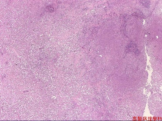
【 Fig. 104-1 (LP)】Necrosis and microabscess formation.
|
|
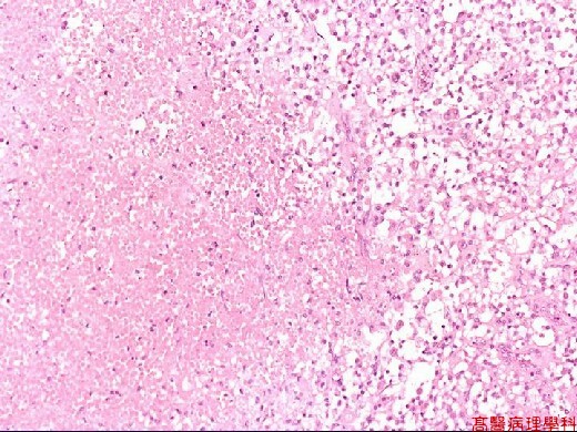
【 Fig. 104-2 (LP)】Necrosis with predominant eosinophils infiltrates.
|
|
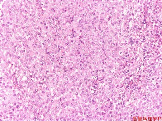
【 Fig. 104-3 (LP)】Proliferation of Langerhans cells with intermingled eosinophils, neutrophils and lymphocytes.
|
|
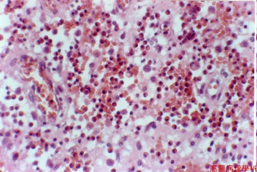
【 Fig. 104-4 (HP)】Eosinophilic microabscess.
|
|

【 Fig. 104-5 (HP)】Langerhans cells: kidney-shaped ,grooved, folded nuclei with vesicular appearance.Fine chromatin and nucleoli are occasionally seen
![]()
![]()
![]()