《Slide 132.》Renal cell carcinoma, Kidney
A. Brief Descriptions:
-
Tumors arise from tubular epithelial cells; Tobacco is the most prominent risk factor; often in older; preponderance in men.
-
Classic symptoms: hematuria, loin pain & mass.
-
Classification of RCC: clear cell (nonpapillary) carcinoma (70% to 80%) ; papillary carcinoma(10% to 15%); chromophobe renal carcinoma (5%).
B. Gross Findings:
-
Protruding from renal cortex, as an irregular, bosselated mass, expansile growth with compressing adjacent renal parenchyma into a pseudocapsule. Cut surface often hemorrhagic, grayish white to yellow, and cystic change or necrosis.
C. Micro Findings:
-
Clear cell:
-
Clear cytoplasm due to high contain of lipid & glycogen.
-
Prominent cell borders and tightly adherent to neighboring cells.
-
-
Granular cell: pink granular cytoplasm due to high content of mitochondria.
-
Arrange in sheets, nests, cords, tubular or papillary pattern with a fine capillary vascular background, and separated by fibrous septi.
-
Little cellular or nuclear pleomorphism or mitosis.
-
Necrosis, fibrosis, cholesterol deposit ( may with foreign body reaction), hemorrhage, calcification or cystic change.
D. Others:
-
Peak at sixth decade, male 2-3 times more frequently than female ; higher incidence in Jews.
E. Reference:
-
Robbins Pathologic Basis of Disease, 6th ed. p.991-994
|
|
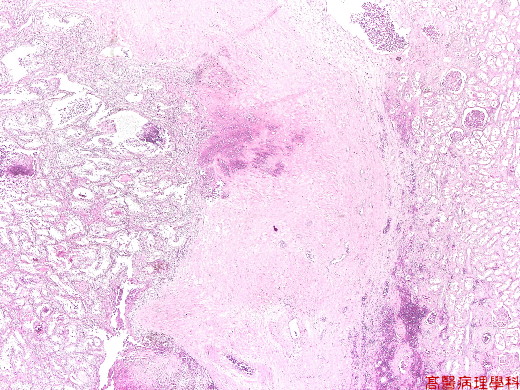
【 Fig. 132-1 (2X)】Tumor mass is seen left field and normal architecture is seen in right view in this scanning field.
|
|
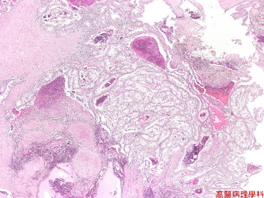
【 Fig. 132-2 (4X)】Tumor exhibits clear appearnce.
|
|
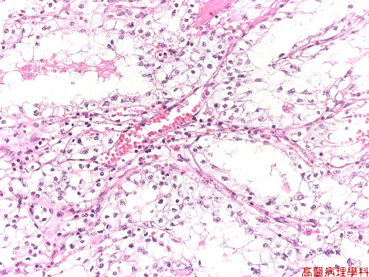
【 Fig. 132-3 (10X)】Clear cytoplasm due to high contain of lipid & glycogen.
|
|
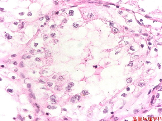
【 Fig. 132-4 (40X)】Clear cytoplasm due to high contain of lipid & glycogen..
![]()
![]()
![]()