《Slide 133.》Squamous cell carcinoma, Kidney
A. Brief Descriptions:
-
Infiltration of tumor cells with a spiky or angular outline which are surrounding by a fibroblastic or fibrous stroma.
-
Related to renal stone.
B. Gross Findings:
略.
C. Micro Findings:
-
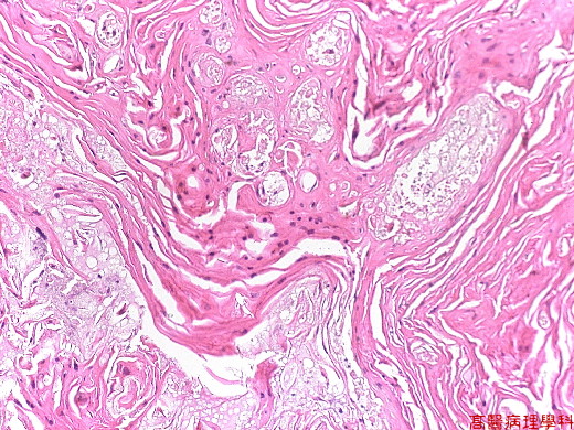
Infiltration of tumor cells with a spiky or angular outline which are surrounding by a fibroblastic or fibrous stroma
-
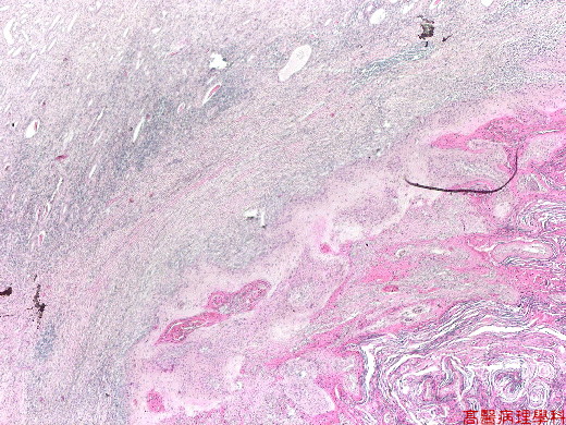
Renal tumor with extensive keratinization (layered eosinophilic materials);note normal renal parenchyma and squamous metaplasia of renal pelvic transitional epithelium
-
Keratinized squamous cancer cells with infiltrating borders; note dilated atrophic ducts
D. Others:
-
Differentiated diagnosis from squamous metaplasia of urothelium (lack of dysplasia) and transitional cell carcinoma with squamous metaplasia.
E. Reference:
-
Robbins Pathologic Basis of Disease, 6th ed. p.262 and p.1006-1007
|
|
【 Fig. 133-1 (2X)】Keratinized squamous cancer cells with infiltrating borders in this scanning view.
|
|
【 Fig. 133-2 (10X)】Tumor cells demonstrating keratinization.
|
|
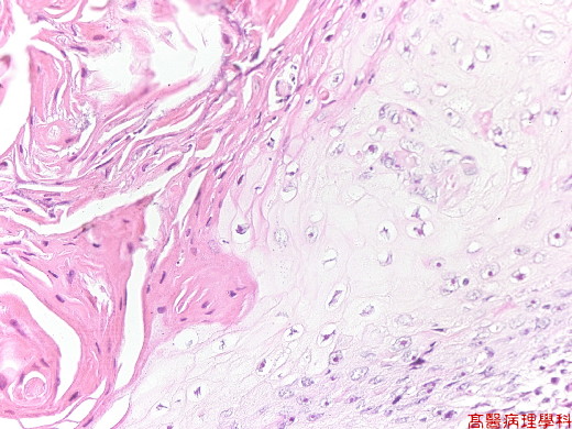
【 Fig. 133-3
(20X)】The
tumor cells show pleomorphism and hyperchromatism.
Keratinization is
seen in left view.
|
|
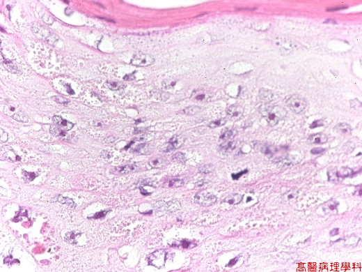
【 Fig. 133-4
(40X)】The tumor cells show pleomorphism and hyperchromatism.
Mitoses is seen in this view.


