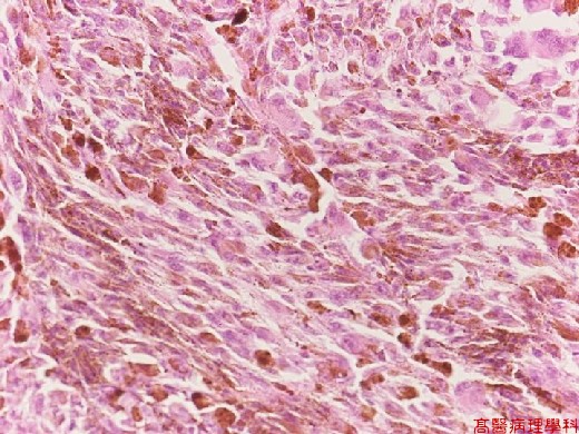
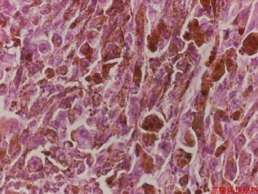
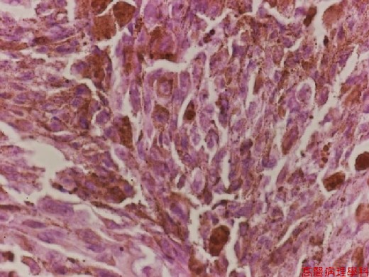
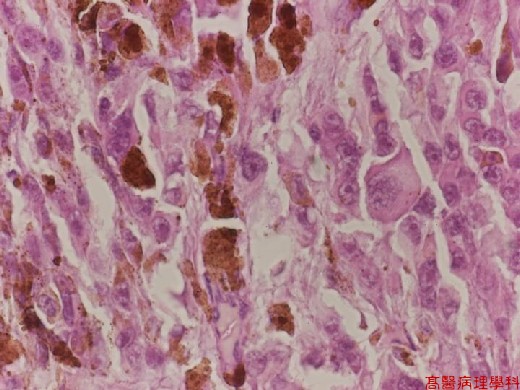
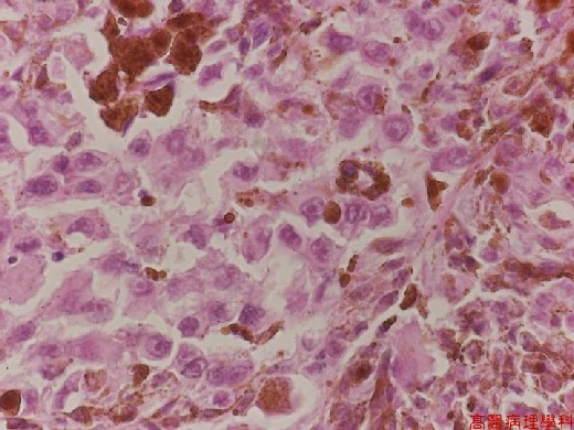
《Slide 97.》Malignant melanoma, Eye
A. Brief Descriptions:
-
Arising from pigmented neuroepithelia or uveal melanocytes.
-
Most common in posterior choroid in eye.
B. Gross Findings:
-
Apple & Blodi classification
-
Obvious and unquestionable nevi and melanocytoma
-
Phase A tumor:
-
Relatively small, not extended or metastatic beyond eye
-
Most cells classified toward benign end and stationary or slow growing
-
-
Phase B tumor:
-
Large, extensive and rapid growth with malignant bizarre cells
-
-
-
Callender classification
-
Spindle A and spindle B cell groups
-
Slender, elongated, spindle shape, cohesive cells
-
With poorly visualized cell border
-
Slender, elongated, spindle shaped nuclei
-
Spindle A cells : devoid of nucleolus
-
Spindle B cells : larger, with prominent nucleoli
-
-
Fascicular: cells in parallel rows, bundles about dilated vessels, with palisading nuclei
-
Mixed spindle and epithelioid cells
-
Epithelioid cells:
-
Larger cells variation in size and shape
-
Well-demarcated, poorly cohesive
-
Abundant cytoplasm, hyperchromatic nucleus with prominent nucleoli
-
Frequent mitosis
-
Multinucleated tumor giant cells
-
-
C. Micro Findings:
-
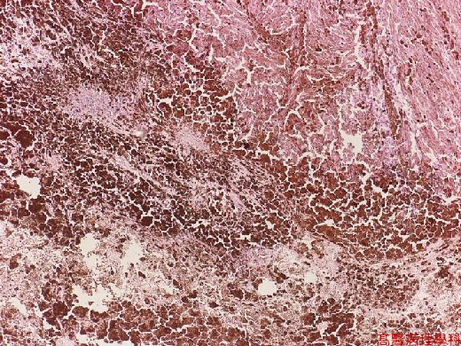
Tumor grows into vitrous cavity with elevated retinal layer with dark-brown melanin pigments.
-
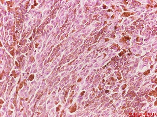
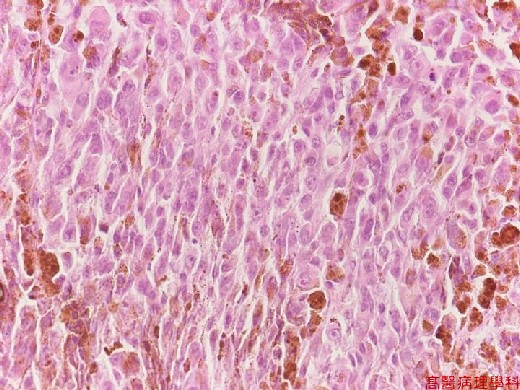
Epithelioid cells with large cell size, abundant cytoplasm, hyperchromatic nuclei and prominent nucleoli.
-
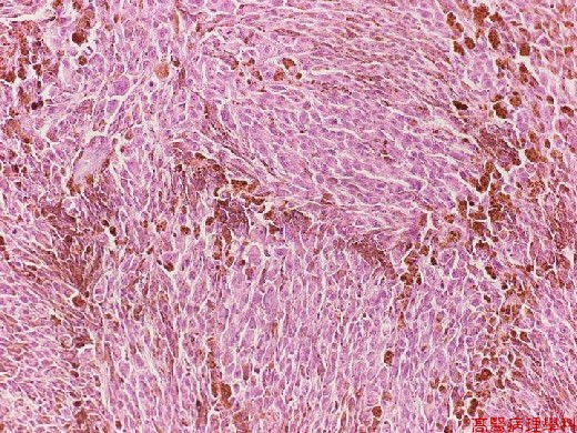
Layer of spindle cells, some with prominent nucleoli.
D. Others:
-
Arising from pigmented neuroepithelia or uveal melanocyte
-
Most common in posterior choroid in eye
E. Reference:
-
Robbins pathologic basis of disease. Ch31. p.1366-1367.
|
|
【 Fig. 97-1 (4X)】
|
|
【 Fig. 97-2 (10X)】
|
|
【 Fig. 97-3 (20X)】
|
|
【 Fig. 97-4 (20X)】
|
|
【 Fig. 97-5 (20X)】
|
|
【 Fig. 97-6 (40X)】
|
|
【 Fig. 97-7 (40X)】
|
|
【 Fig. 97-8 (40X)】
|
|
【 Fig. 97-9 (40X)】
