《Slide
155.》Giant
cell tumor,
Bone
A. Brief Descriptions:
-
Most cases older than 19 years at the time of diagnosis.
-
The distal femur and proximal tibia account for 60% of cases.
-
Swelling in the region of affected bone with pain.
B. Gross Findings:
-
Gray-brown to reddish tumor with necrosis and cyst formation
C. Micro Findings:
-
A uniform pattern of growth on low magnification.
-
Multinucleated giant cells lie scattered uniformly in a “sea” of mononuclear round to oval cells.
-
The nuclear of mononucleated cells and those of the multinucleated giant cells are similar in appearance and uniform in their cytological characteristics.
-
Mitotic figures are present in some mononuclear cells but absent in the giant cells.
-
Hemorrhage and degeneration are common.
D. Others:
略.
E. Reference:
-
Robbins Pathologic Basis of Disease, 6th ed. P1244-1245.
|
|
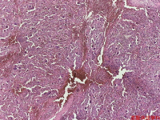
【 Fig. 155-1 (LP)】Cellular tumor with hemorrhage.
|
|
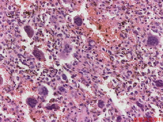
【 Fig. 155-2 (LP)】Numerous multinucleated giant cells lie scattered in a “sea” of mononuclear round to oval cells.
|
|
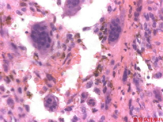
【 Fig. 155-3 (HP
![]()
![]()
![]()