《Slide
158.》Osteosarcoma,
Femur
A. Brief Descriptions:
-
20% of primary bone cancers.
-
Usually arise in the metaphyseal region of the long bones of the extremities.
-
Radiographically it reveals Codman’s triangle and bony spiculus (sun ray appearance).
B. Gross Findings:
-
Gritty or gray-white.
-
Contain areas of hemorrhage and cystic degeneration.
C. Micro Findings:
-
Osteoid.
-
The tumor cells vary in size and shape and frequently have large hyperchromatic nuclei.
-
Wovening bone formation.
-
Bizarre tumor giant cells are common, as are mitoses.
D. Others:
略.
E. Reference:
-
Robbins Pathologic Basis of Disease, 6th ed. P1236-1237.
|
|
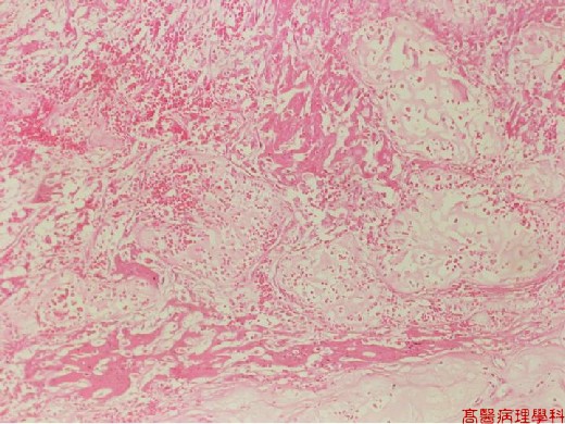
【 Fig. 158-1 (LP)】 A network of thin and broader trabeculae of osteoid.
|
|
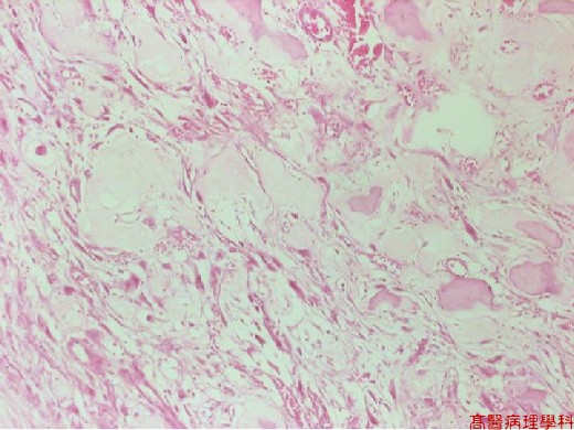
【 Fig. 158-2 (LP)】
|
|
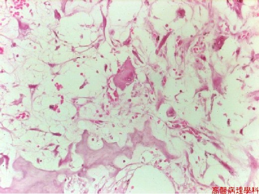
【 Fig. 158-3 (HP)】The tumor cells vary in size and shape and frequently have large hyperchromatic nuclei.
![]()
![]()
![]()