Slide 41.Amebiasis, Colon

A. Brief Descriptions

  1. Caused by the protozoan parasite Entamoeba histiolytica.

  2. Infections from---E. histiolytica cysts (achitin wall & 4 nuclei).                                       

  3. Ameboid from----trophozoites.

  4. Most frequently involves the cecum and ascending colon followed by sigmoid à rectum à appendix.

B. Gross Findings

      略.

C. Micro Findings

  1. Ulcerated mucosa, edema, congestion and a prominent infiltrate of eosinophils.

  2. Round organisms of 20-40 um diameter, some ingested RBCs (erythrophagocytosis ), are found in loose surface debris.

D. Others:

  1. Diagnosis is best made by identified hematophagous trophozoites in fresh stools.

E. Reference

  1. Robbins Pathologic Basis of Disease, 6th ed.  P.358-359.
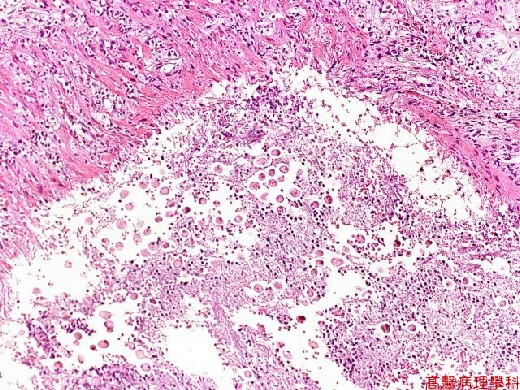


Fig. 41-1 (LP)Numerous eosinophilic spherical structure within necrotic area.
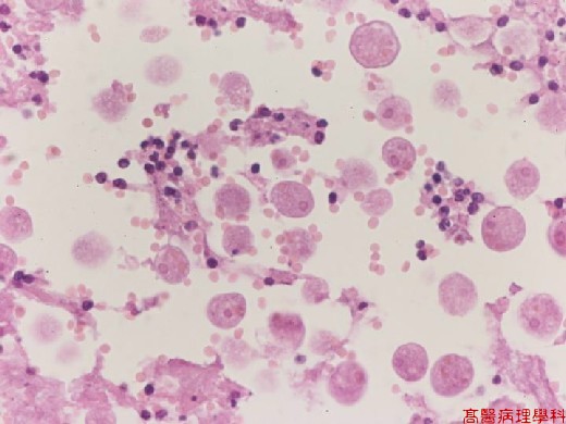


Fig. 41-2 (HP)The spherical structure (trophozoites) has one basophilic

                                                       nuclei about the size of RBC’s. Note some RBCs are

                                                       phagocytized by the trophozoites (erythrophagocytosis).