《Slide 44.》Pneumocystis carinii pneumonia & Cytomegalovirus (CMV) pneumonitis, Lung
A. Brief Descriptions:
-
Pnumocystis carinii pneumonia:
(1) Cup-shaped or boat-shaped cysts (4~6 mm).
(2) Diffuse or patchy pneumonia.
(3) The affected lung are airless, red and beefy.
-
CMV pneumonitis: interstitial pneumonitis.
B. Gross Findings:
-
PCP : Airless, red, or beefy lung.
C. Micro Findings:
Pneumocystis carinii pneumonia
HE: the alveolar spaces filled by an amphophilic, foamy, amorphous material resembling proteinaceous edema fluid composed of proliferating parasites & cell debris.
Toluidine blue: trophozoites.
Methenamine-silver: typical cup-shaped cyst with trophozoites in the cyst.
Cytomegalovirus (CMV) pneumonitis
Interstitial pneumonitis by the characteristic intranuclear inclusion body surrounded by a clear halo in the enlarged alveolar lining cells, endothelial cells of septal capillaries & alveolar macrophages.
D. Others:
Pneumocystis carinii pneumonia
-
Opportunistic infection of pnemocystis carinii, measuring up to 6 um, usually concurrent infection by opportunistic bacteria, fungi, viruses especially CMV.
Cytomegalovirus (CMV) pneumonitis
-
Opportunistic infection in debilitated or immunocompromised patient, e.g. AIDS.
E. Reference:
-
Robbins Pathologic Basis of Disease, 6th ed. P.376,381-382.
|
|
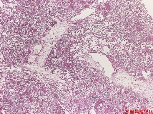
【 Fig. 44-1 (LP)】A high proportion of alveolar space is filled with pinkish substance, so the airspace is obliterated, and the architecture of lung is hard to identified at a glance.
|
|
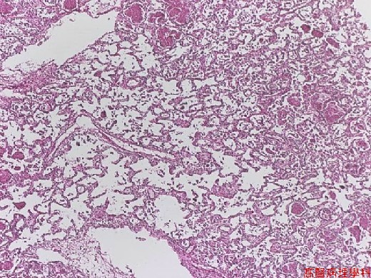
【 Fig. 44-2 (LP)】The non-involved alveoli show emphysematous change.
|
|
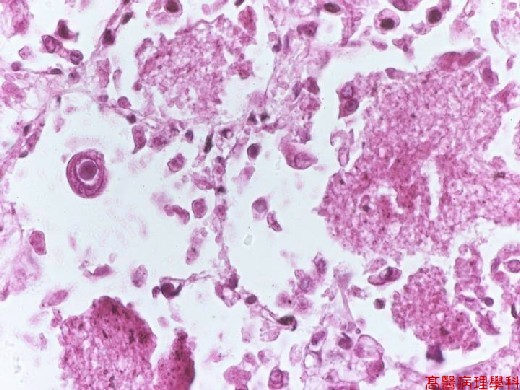
【 Fig. 44-3 (HP)】The alveolar space is filled with foamy amorphous material ( right, upper and lower). A CMV-infected enlarged epithelial cell with typical bull-eye appearance with large intranuclear inclusion body surrounded by a clear halo.
|
|
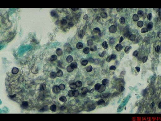
【 Fig. 44-4 (HP, silver stain)】The cup-shaped Pneumocystis carinii organisms within a sputum sample.
![]()
![]()
![]()