《Slide 72.》Cystic mature teratoma, Testis
A. Brief Descriptions:
-
The tumor is composed of neural tissue,squamous epithelium and connective tissue stroma.
-
Devoid of EC cells.
B. Gross Findings:
Cystic & multiloculated.
C. Micro Findings:
-
All tissue are well differentiated or matured.
-
All types of tissue can be seen, the most common being nerve, cartilage, smooth muscle & various types of epithelium (alimentary, respiratory, squamous.
D. Others:
-
Definition:true neoplasm arising from totipotential cells & composed therefore of numerous types of tissue (more than one germinal layer).
-
Mature teratoma of testis:the least aggressive of the nonseminomatous germ cell tumors of the testis.
E. Reference:
Robbins Pathologic Basis of Disease, 6th ed. P.1021-1022.
|
|
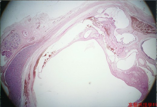
【 Fig. 72-1 (1X)】All types of tissue can be seen.
|
|
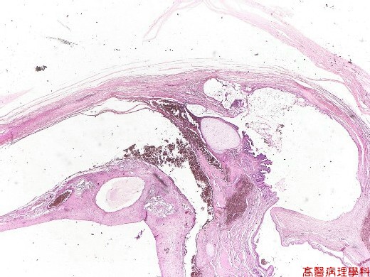
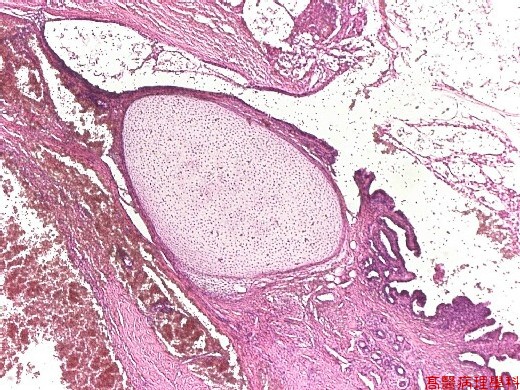
【 Fig. 72-2 (2X)】Lobulated and cystic lesion with cartilage seen in this view.
|
|
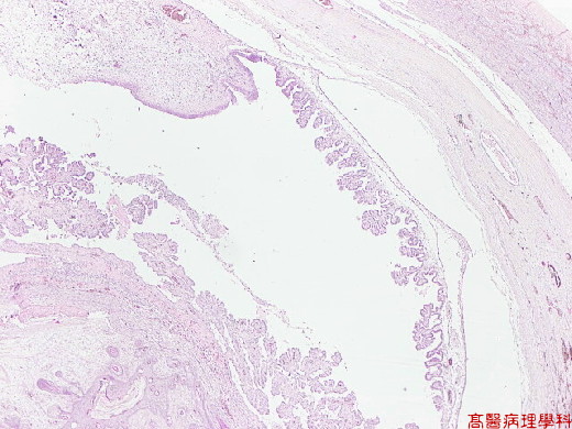
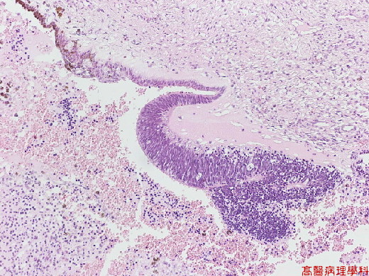
【 Fig. 72-3 (2X)】Choroid plexus-like structure seen in this view.
|
|
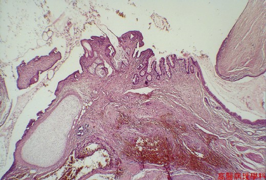
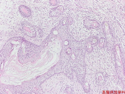
【 Fig. 72-4 (4X)】Skin elements and cartilage seen in this view.
|
|
【 Fig. 72-5 (10X)】Mature cartilage seen in this picture.
|
|
【 Fig. 72-6 (4X)】Skin element with hair shafts seen.
|
|
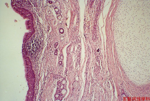
【 Fig. 72-7 (4X)】Ciliated epithelium and glands seen in lefrt view.
|
|
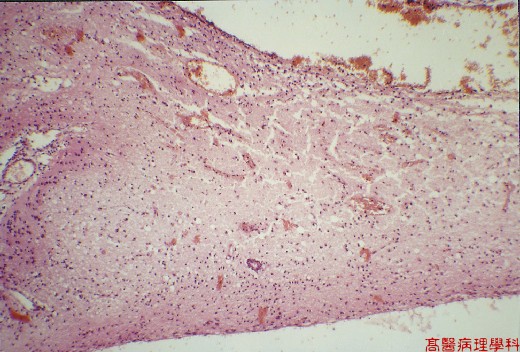
【 Fig. 72-8 (4X)】Brain tissue seen in this picture.
|
|
【 Fig. 72-9 (10X)】Epithelial component seen.
|
|
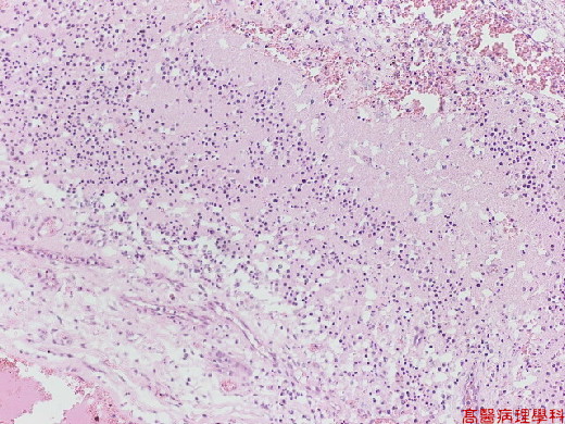
【 Fig. 72-10 (10X)】Brain tissue seen in this picture.
|
|
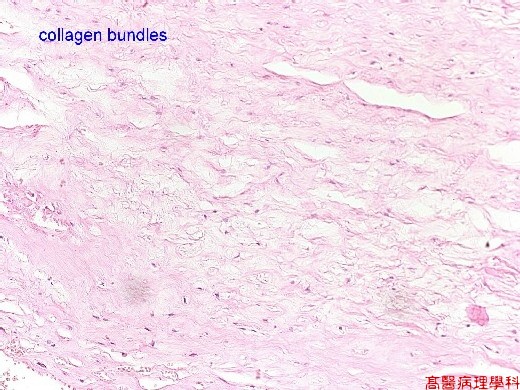
【 Fig. 72-11 (10X)】Collagen seen in this view.
|
|
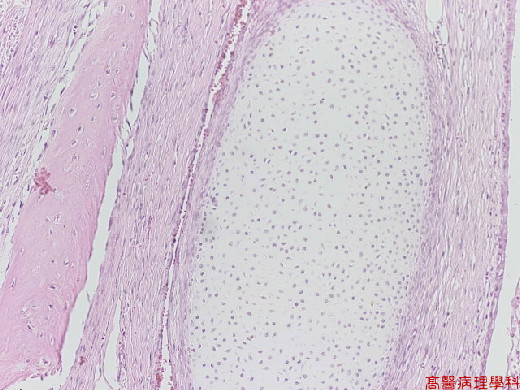
【 Fig. 72-12 (10X)】Bone and cartilage are seen.
|
|
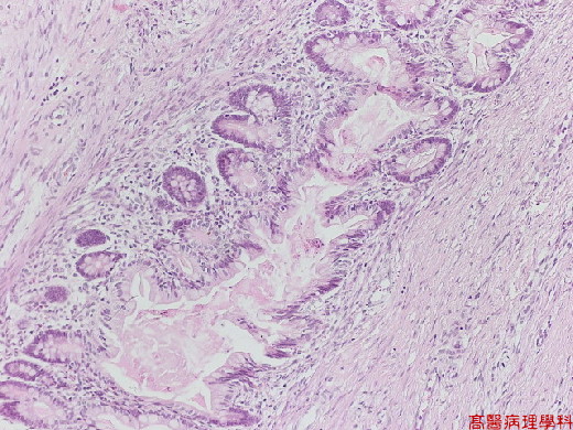
【 Fig. 72-13 (10X)】Gastrointestinal mucosa seen in this view.
![]()
![]()
![]()