《Slide 86.》Intraductal carcinoma, Breast
A. Brief Descriptions:
-
Ductal carcinoma in situ.
-
Definition: Ductal carcinoma with intact basement membrane.
-
Five architectural subtypes: comedocarcinoma, solid, cribriform, papillary, and micropapillary.
B. Gross Findings:
-
Poorly defined focus of slightly increased consistency
-
Punctate areas of necrotic material (comedone-like)
C. Micro Findings:
-
Dilated ducts filled with neoplastic anaplastic epithelial cells that plug lumina.
-
Presence of central necrosis in some ducts.
-
Focal cribriform growth pattern.
-
Tumor cells: loosely cohesive, cytologic atypia, increased mitotic figures.
D. Others:
- Compare with slide 87, infiltrating ductal carcinoma of breast.
E. Reference:
-
Robbins Pathologic Basis of Disease, 6th ed. P.1107-1109.
|
|
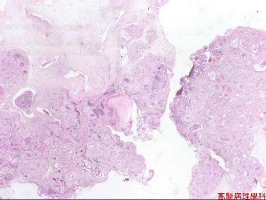
【 Fig. 86-1 (LP)】Poorly defined tumor mass with focal necrosis.
|
|
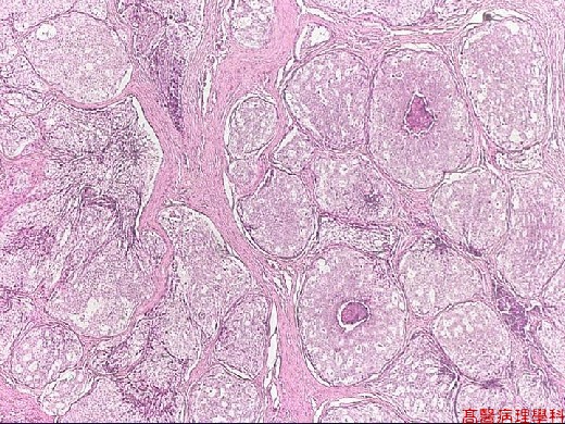
【 Fig. 86-2 (LP)】Dilated ducts with intact basement membrane and filled with neoplastic epithelial cells.
|
|
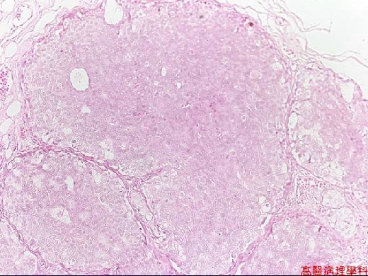
【 Fig. 86-3 (LP)】Solid pattern.
|
|

【 Fig. 86-4 (LP)】Cribriform pattern.
|
|
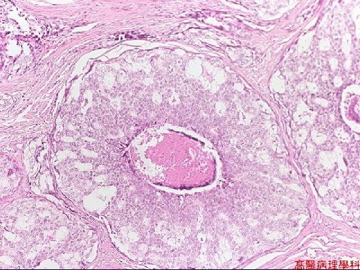
【 Fig. 86-5 (LP)】Comedocarcinoma showing intraductal proliferation of malignant cells with central necrosis.
|
|
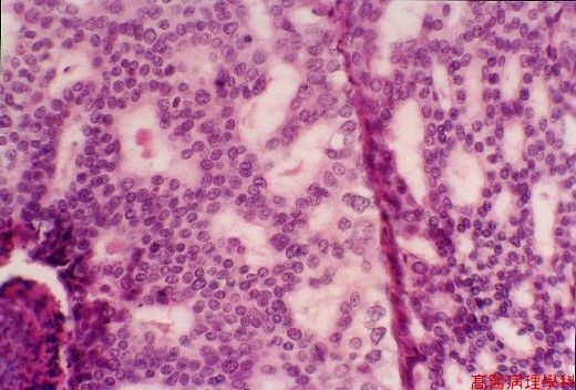
【 Fig. 86-6 (HP)】The tumor cells showing monomorphic appearance and lacking normal myoepithelial cells.Cellular atypia and loose cohesion are noted.
![]()
![]()
![]()