《Slide 117.》Cirrhosis (postnecrotic), Liver
A. Brief Descriptions:
-
Mainly due to chronic viral hepatitis.
-
Irregular size of nodules.
B. Gross Findings:
-
Fibrosis, regenerated nodules & complete vascular distortion of the liver.
C. Micro Findings:
-
Nodules of varying size.
-
Thick or thin fibrous septi.
-
Inflammatory cells, bile ducts, vessels within the fibrous septi.
D. Others:
略.
E. Reference:
-
Robbins Pathologic Basis of Disease, 6th ed. P.864~866.
|
|

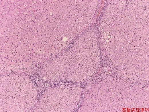
【 Fig. 117-1 (LP)】The entire liver parenchyma is divided into nodules of variable size.
|
|
【 Fig. 117-2 (LP)】Dense inflammatory infiltrate in the fibrous septae segregated liver parenchyma.
|
|
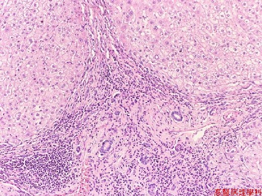
【 Fig. 117-3 (LP)】Widening and fibrosis of periportal area connecting with each other resulting in nodules formation. Note dense inflammatory cells infiltrate in fibrotic bands.
|
|

【 Fig. 117-4 (LP)】Areas of fatty metamorphosis of hepatocytes are also seen.
|
|
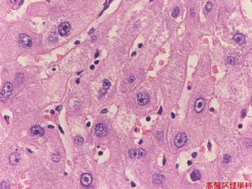
【 Fig. 117-5 (LP)】Regeneration nodules showing regenerating atypia of hepatocytes (multinucleation and hyperchromatism).
![]()
![]()
![]()