《Slide 118.》Hepatocellular carcinoma, Liver
A. Brief Descriptions:
-
Strongly linked to prevalence of HBV infection.
-
Usually paler than the surrounding liver substance.
-
Necrosis, hemorrhage, and bile stained may seen grossly.
B. Gross Findings:
-
Well demarcated nodules with necrosis, hemorrhage & bile stained; soft bulging cut surface.
C. Micro Findings:
-
Tumor cells have eosinophilic cytoplasm & round, vesicular nuclei with distinct nucleoli.
-
Arranged in plates or trabeculae, usually several cell thick, & separated by sinusoidal channels which have an endothelial & very scanty reticulin, but no Kupffer cells.
-
Or arranged in acinar pattern & solid pattern.
-
Granular or clear cytoplasm (glycogen in cytoplasm).
-
Sometimes pleomorphic or multinucleated giant tumor cells.
-
Vascular invasion.
D. Others:
-
Alfa-fetoprotein is of great diagnostic value.
E. Reference:
-
Robbins Pathologic Basis of Disease, 6th ed. P.888~890.
|
|
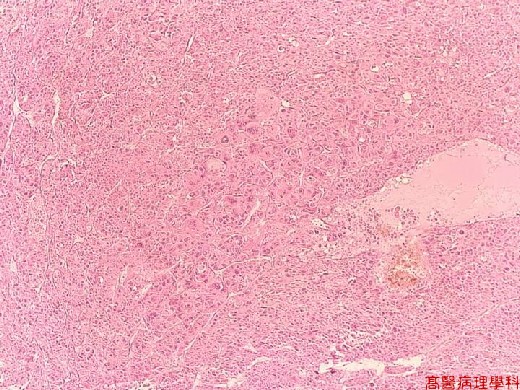
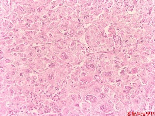
【 Fig. 118-1 (LP)】Nearly solid growth pattern replaced the normal hepatic cord architecture.
|
|
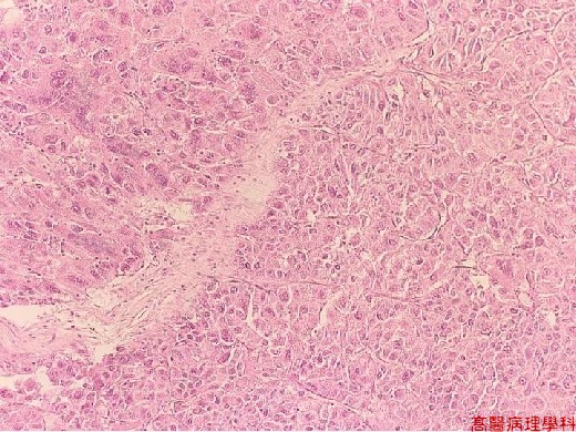
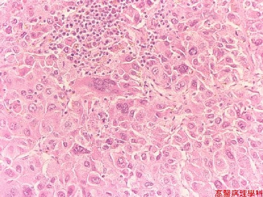
【 Fig. 118-2 (LP)】Small solid nesty architecture is seen in this field. Note the conspicuous cellular atypia.
|
|
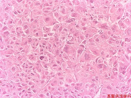
【 Fig. 118-3 (HP)】Cancer cells with hyperchromatism, pleomorphism and high N/C ratio.
|
|
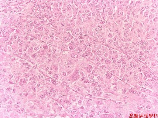
【 Fig. 118-4 (HP)】In this picture, thick trabecular arrangement of cancer cells is seen. Note a mitotic figure in upper middle field.
|
|
【 Fig. 118-5 (HP)】Acinar-like arrangement of cancer cells.
|
|
【 Fig. 118-6 (LP)】Bizarre multinucleated cancer giant cells are not infrequently seen.
![]()
![]()
![]()