《Slide 171.》Cholesterolosis, Gallbladder
A. Brief Descriptions:
-
Mucosal surface is studded with yellow fleck.
-
Not directly related to gallstone formation.
B. Gross Findings:
略.
C. Micro Findings:
-
Foamy cells aggregate in mucosa.
D. Others:
略.
E. Reference:
-
Robbins Pathologic Basis of Disease, 6th ed. P.895.
|
|
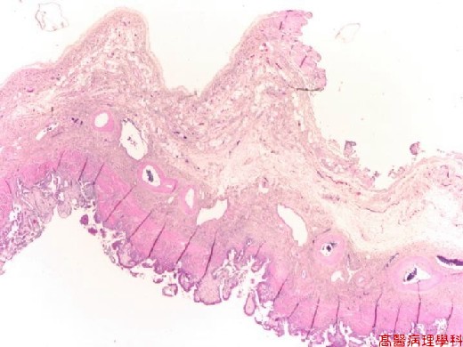
【 Fig. 171-1 (LP)】Randomly distributed pale areas between mucosa and muscle layer.
|
|
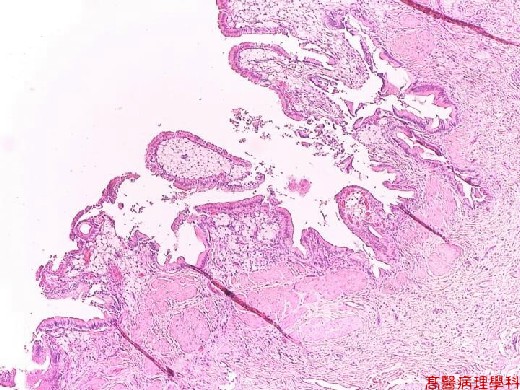
【 Fig. 171-2 (LP)】Accumulation of foamy histiocytes in lamina propria.
|
|

【 Fig. 171-3 (HP)】Uniform round to polyhedral foamy histiocytes with centrally located small round nuclei with very low N/C ratio aggregate in lamina propria just beneath surface epithelium.
![]()
![]()
![]()