《Slide 179.》Cholangiocarcinoma, Liver
A. Brief Descriptions:
-
Less common than hepatocellular carcinoma
-
No direct relationship with cirrhosis
-
Resemble adenocarcinoma arising in the other part of the body.
-
Rare bile stained.
B. Gross Findings:
略.
C. Micro Findings:
-
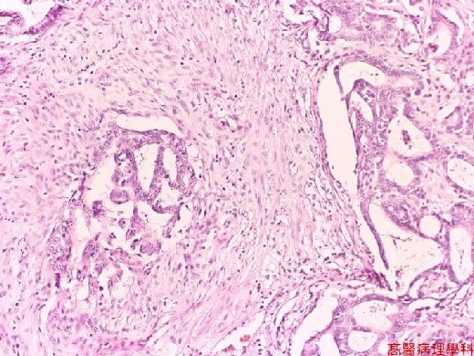
Clearly defined neoplastic glandular and tubular structures in portal area.
-
Lined by anaplastic cuboid to low columnar epithelial cells.
-
Markedly desmoplastic stroma.
D. Others:
略.
E. Reference:
-
Robbins Pathologic Basis of Disease, 6th ed. P.888-890.
|
|
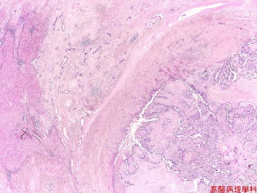
【 Fig. 179-1 (LP)】The hepatic parenchyma is infiltrated by irregular glandular or papillary structures. The normal liver is in the left.
|
|
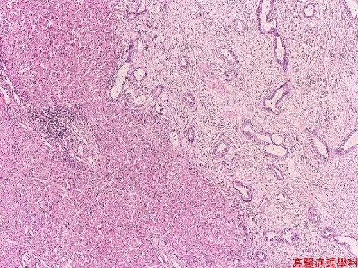
【 Fig. 179-2 (LP)】The irregular cancerous glands infiltrate in liver parenchyma.
|
|
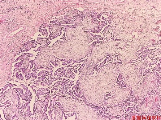
【 Fig. 179-3 (LP)】Marked stromal desmoplasia. The fibrotic tissue surrounds cancerous glands.
|
|
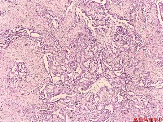
【 Fig. 179-4 (LP)】The cancer cells form ill-defined or fused glandular structure.
|
|
【 Fig. 179-5 (LP)】The hyperchromatic, pleomorphic cancer cells arrange in irregular glandular or cribriform patterns.
![]()
![]()
![]()