《Slide 58.》Tubulovillous adenoma, Colon
A. Brief Descriptions:
-
The prevalence of colonic adenomas is about 20 to 30 % before age of 40, arising to 40 to 50% after age of 60.
-
Adeomatous polyps are segregated into 3 subtypes on the basis of epithelial architecture.
-
Tubular adenoma: exhibit greater than 75% tubular architecture.
-
Villous adenoma: contain greater than 50% villous architecture.
-
Tubulovillous adenoma: contain 25 to 50% villous architecture.
B. Gross Findings:
-
Range from small, pedunculated lesions to large neoplasm that are usually sessil.
C. Micro Findings:
-
Neoplastic (adenomatous) epithelium: enlarged nuclei, hyperchromatic and elongated in shape; stratification of nuclei; varies degree of dysplasia.
-
Tubular adenoma: proliferation of adenomatous epithelium, that forms tubules (separated from each other by normal lamina propria), regular tubules with little branching or tufting.
-
Villous adenoma: growth of fine fingerlets or villi that project perpendicularly from the muscularis mucosa to the outer tip of the adenoma.
D. Others:
-
All adenomatous lesions arise as the result of epithelial proliferative dysplasia, which may range from mild to severe (as carcinoma in situ).
-
There is strong evidence that adenoma are precursor lesion for invasive colorectal adenocarcinoma.
E. Reference:
-
Robbins Pathologic Basis of Disease, 6th ed. P.827 ~ 831.
|
|
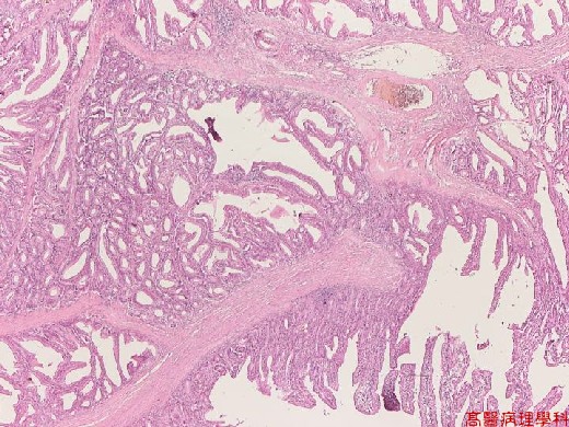
【 Fig. 58-1 (LP)】The tumor compose of tubular and villous architecture.
|
|
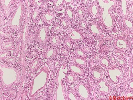
【 Fig. 58-2 (LP)】Tubular adenoma: proliferation of adenomatous epithelium, that forms tubules (separated from each other by normal lamina propria), regular tubules with little branching or tufting.
|
|
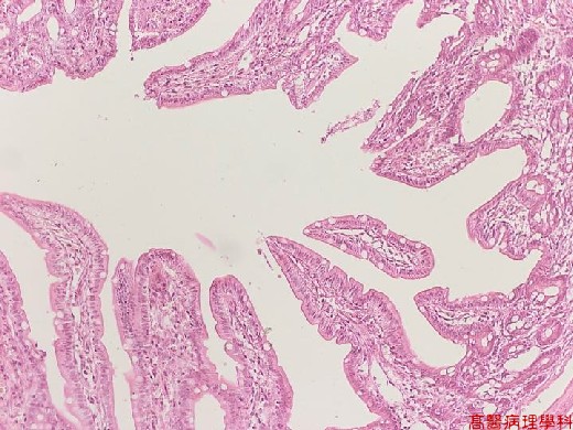
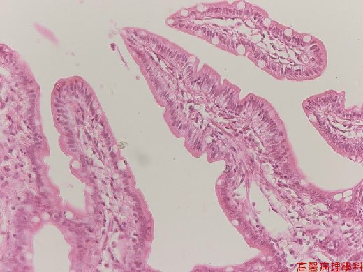
【 Fig. 58-3 (LP)】Villous adenoma: growth of fine fingerlets or villi that project perpendicularly from the muscularis mucosa to the outer tip of the adenoma.
|
|
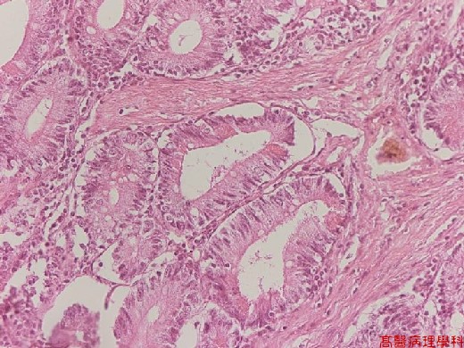
【 Fig. 58-4 (HP)】Neoplastic (adenomatous) epithelium: enlarged nuclei, hyperchromatic and elongated in shape; stratification of nuclei; varies degree of dysplasia.
|
|
【 Fig. 58-5 (HP)】Neoplastic (adenomatous) epithelium: enlarged nuclei, hyperchromatic and elongated in shape; stratification of nuclei; varies degree of dysplasia.
![]()
![]()
![]()