《Slide 66.》Carcinoid tumor, Stomach
A. Brief Descriptions:
-
In the mucosa of the G-I tract, there are scattered endocrine cells which synthesize, store, and secret biogenic amines or peptides participating in coordinate gut function. The tumor arise from these cells are designated as carcinoid tumor.
-
The appendix is the most common site of gut carcinoid tumor, followed by the small intestine, rectum, stomach and ileum.
-
Carcinoid tumor are potentially malignant and the tendency of malignant behavior correlate with the site of origin, the depth of local penetration and the size of the tumor.
B. Gross Findings:
-
The tumor appear as intramural or submucosal masses that creat small, polypod or plateau-like elevations rarely more than 3 cm in diameter.
-
The overlying mucosa may be intact or ulcerated.
C. Micro Findings:
-
Monomorphic cells in trabecular, solid, insular, festoon (ribbon-like), or tubular structures, some small glands with a rosette-like appearance.
-
Tumor cells have uniform round to oval nuclei and scant to moderate amount of finely granular cytoplasm.
-
Central located nuclei with prominent nucleoli and fine chromatin (salt & pepper).
-
Rich of vascularity and prominent fibrous stroma.
-
Atypia may be seen.
D. Others:
略.
E. Reference:
-
Robbins Pathologic Basis of Disease, 6th ed. P.835 ~ 837.
|
|
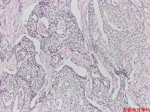
【 Fig. 66-1 (LP)】The tumor cells arrange in solid nests or trabeculae with glandular formation in the fibrovascular stroma.
|
|
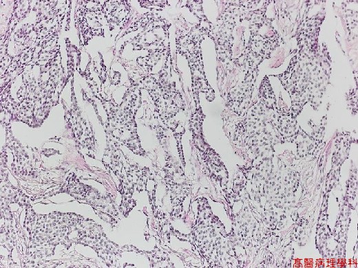
【 Fig. 66-2 (LP)】Insular, or festoon (ribbon-like) structure.
|
|
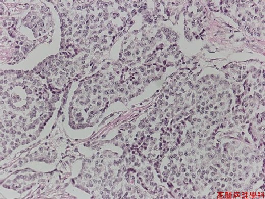
【 Fig. 66-3 (HP)】The tumor cells are monotonously similar, having a scant, pink granular cytoplasm.
|
|
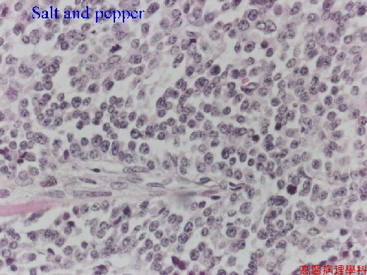
【 Fig. 66-4 (HP)】Central located nuclei with prominent nucleoli and fine chromatin (salt & pepper appearance).
![]()
![]()
![]()