《Slide 139.》Seborrheic keratosis, Skin
A. Brief Descriptions:
1. Single but often multiple.
2. Middle age, on the trunk, face, and extremities.
3. Smooth surface with keratotic plugs.
B. Gross Findings:
-
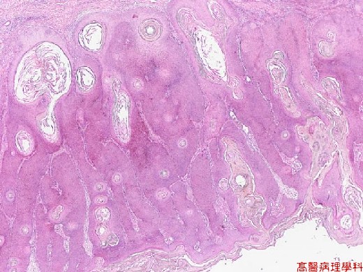
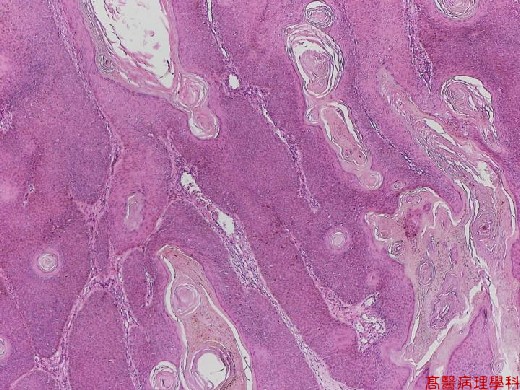
Sharply demarcated, brownish in color, and slightly raised, most of them have a verrucous surface
C. Micro Findings:
-
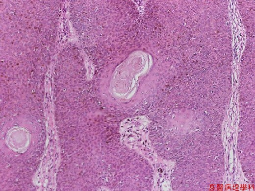
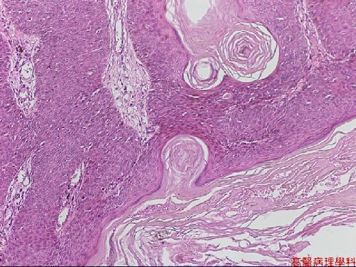
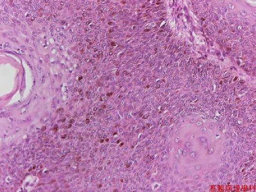
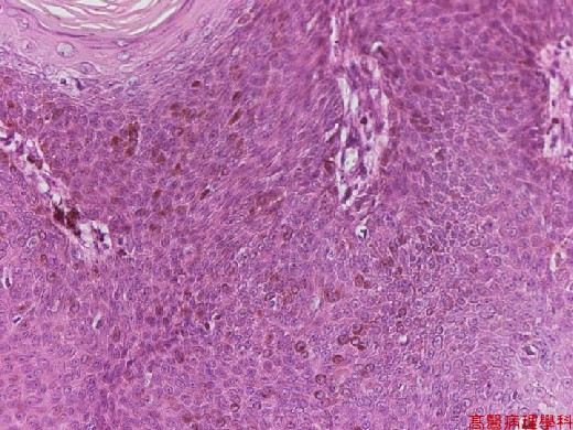
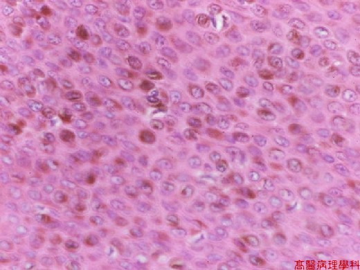
Exophytic and composed of basaloid cells.
-
The base of the seborrheic keratosis is flat and level with the base of the adjacent normal epidermis.
-
The basaloid cells (same appearance of normal epidermal basal cells) form cords and sheets in the middle of which are epidermal cysts filled with keratin (horn cyst), some keratotic cysts communicate with the epidermal surface (pseudohorn cysts).
-
Hyperpigmentation of the basaloid cells may be present.
D. Others:
-
Benign tumor of basaloid (basal cell-like) cells.
-
Predilection site: trunk and face.
-
Age: after middle age.
-
Leser-Trelat sign: sudden appearance of numerous seborrheic keratoses in association with a malignant tumor.
E. Reference:
-
Robbins Pathologic Basis of Disease. Ch27. p.1180.
|
|
【 Fig. 139-1 (2X)】
|
|
【 Fig. 139-2 (4X)】
|
|
【 Fig. 139-3 (10X)】
|
|
【 Fig. 139-4 (10X)】
|
|
【 Fig. 139-5 (20X)】
|
|
【 Fig. 139-6 (20X)】
|
|
【 Fig. 139-7 (40X)】
![]()
![]()
![]()