《Slide 140.》Keratoacanthoma, Skin
A. Brief Descriptions:
1. Rapidly developing neoplasm.
2. Sun exposed skin of whites older than 50 years.
3. Flesh-colored, dome-shaped nodules with a central keratin-filled plug. (crater-like)
B. Gross Findings:
-
Begins as a firm, round, flesh colored or red papule later forming a central crater filled with keratin
C. Micro Findings:
-
Large, central keratin-filled crater.
-
The epidermis extends like a lip over the sides of the crater.
-
At the base of the crater, irregular epidermis proliferation extend both upward into the crater and downward from the base of the crater.
-
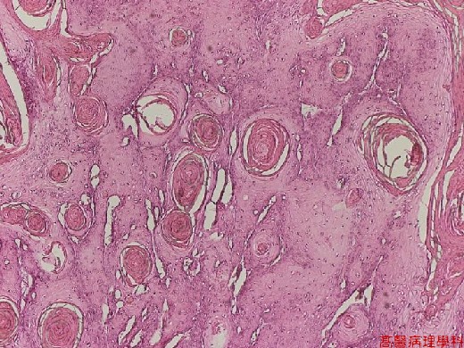
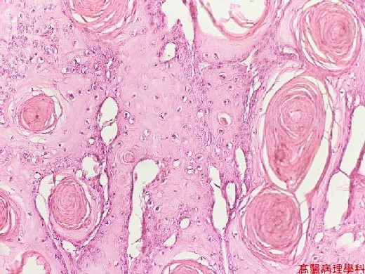
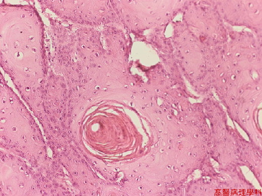
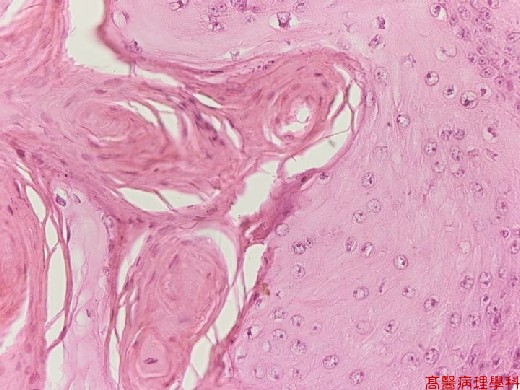
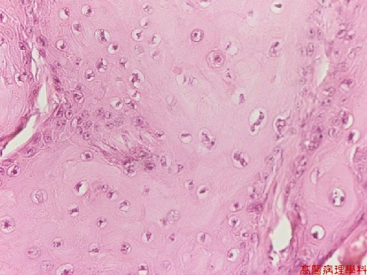
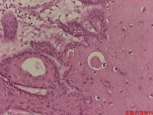
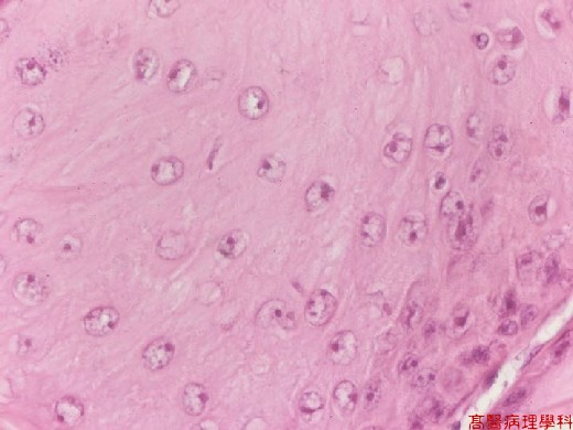
In more centrally of the epidermal proliferation large squamoid cells have a characteristic homogenous and pale-pink (" glassy ") cytoplasm; at the periphery, the cells are basaloid.
-
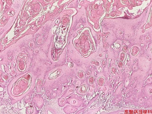
There are many horn pearls, most of which shows complete keratinization in their center.
-
The base of fully developed keratoacanthoma appears regular and well demarcated.
-
Rather dense inflammatory infiltrate is present at the base of the lesion.
D. Others:
-
Benign but rapidly growing tumor
-
Predilection site: central face
-
Age: at or after middle age
E. Reference:
-
Robbins Pathologic Basis of Disease. Ch27. p.1181
|
|
【 Fig. 140-1 (4x)】
|
|
【 Fig. 140-2 (10X)】
|
|
【 Fig. 140-3 (20X)】
|
|
【 Fig. 140-3 (20X)】
|
|
【 Fig. 140-4 (20X)】
|
|
【 Fig. 140-5 (20X)】
|
|
【 Fig. 140-6 (20X)】
|
|
【 Fig. 140-7 (40X)】
