《Slide 141.》Bowen's disease, Skin
A. Brief Descriptions:
1. Occur on exposed or on unexposed skin and in pigmented or poorly pigmented skin.
2. Intraepidermal squamous cell carcinoma, squamous cell carcinoma in situ.
3. Slightly elevated, scaly, flesh-colored plaque of long duration.
B. Gross Findings:
-
Slowly enlarging erythematous patch of sharp but irregular outline; within the patch are general areas of scaling and crusting
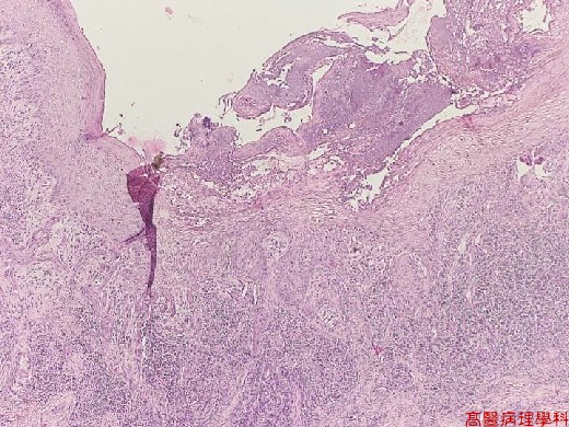
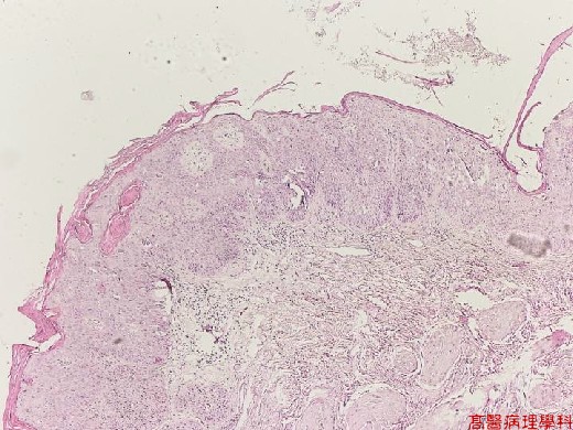
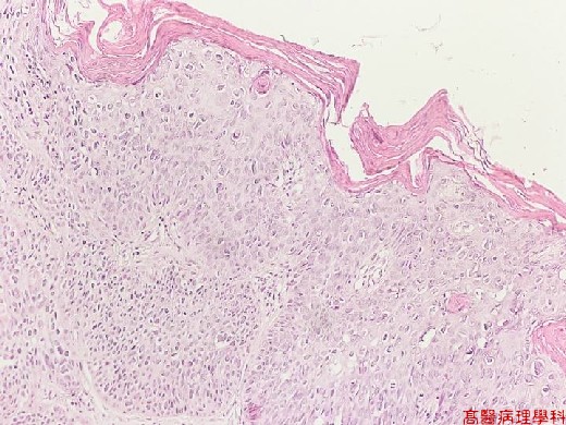
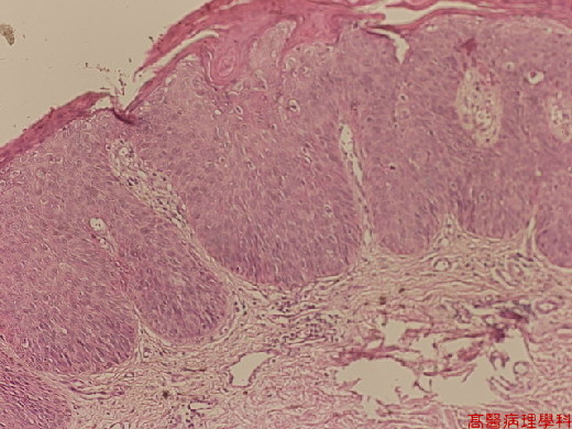
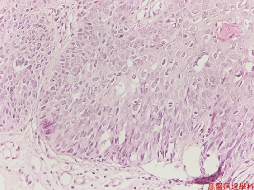
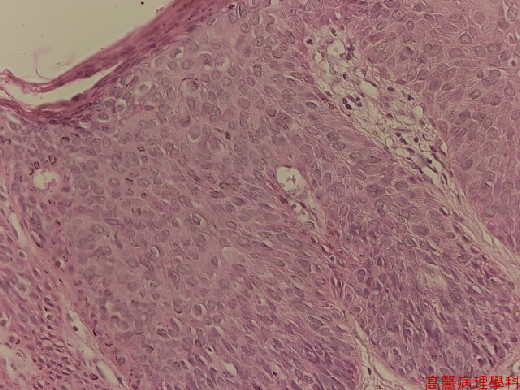
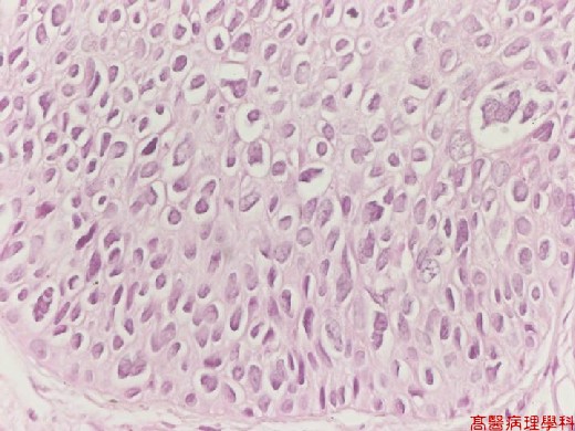
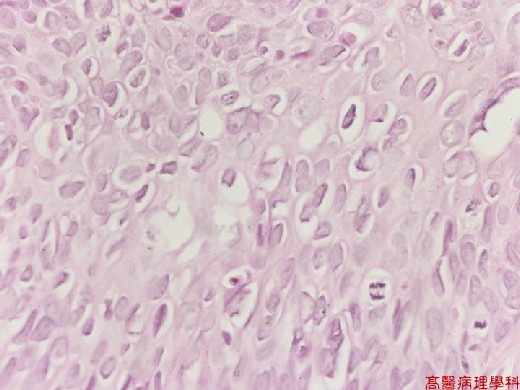
C. Micro Findings:
-
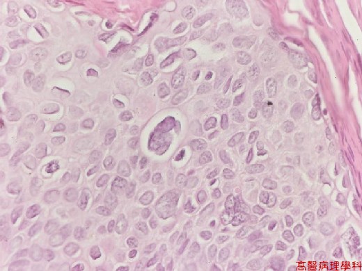
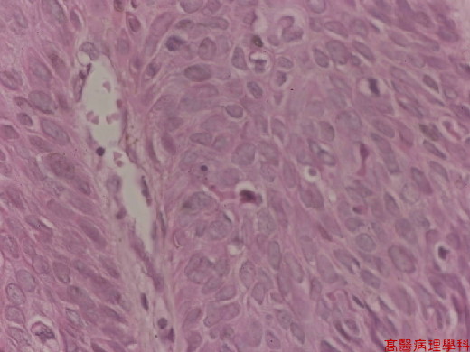
The epidermis is thickened.
-
The border between the epidermis and dermis appears sharp.
-
Throughout the epidermis, cells lie in complete disorder, and many of the them appear atypical, showing large, hyperchromatic nuclei.
-
In the epidermis there are scattered cells showing atypical individual cells’ keratinization (dyskeratosis).
-
The horny layer usually is thickened and consists largely of parakeratotic cells with atypical, hyperchromatic nuclei.
D. Others:
略
E. Reference:
略
|
|
【 Fig. 141-1 (4X)】
|
|
【 Fig. 141-2 (4X)】
|
|
【 Fig. 141-3 (10X)】
|
|
【 Fig. 141-4 (10X)】
|
|
【 Fig. 141-5 (20X)】
|
|
【 Fig. 141-6 (20X)】
|
|
【 Fig. 141-7 (40X)】
|
|
【 Fig. 141-8 (40X)】
|
|
【 Fig. 141-9 (40X)】
|
|
【 Fig. 141-10 (40X)】
![]()
![]()
![]()