《Slide
36.》Pleomorphic
adenoma (mixed tumor),
Salivary gland
A. Brief Descriptions:
-
The most common tumor, according for 60 percent to 70 percent of all parotid tumors and for 40 percent to 70 percent of tumors in other glands.
-
Benign tumor; consisting of cells with epithelial(luminal) and myoepithelial (abluminal) differentiation, and variable amounts of characteristic stroma.
-
Have also been called mixed tumor.
B. Gross Findings:
Rubbery, resilient mass with a bosselated surface.
C. Micro Findings:
-
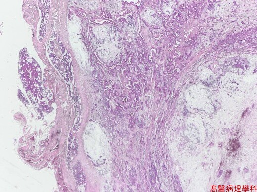
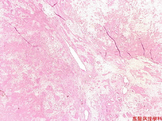
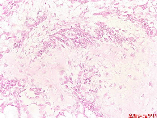
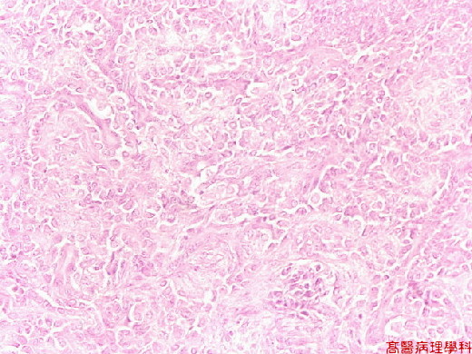
Biphasic appearance resulting from the admixture or epithelial & stroma.
-
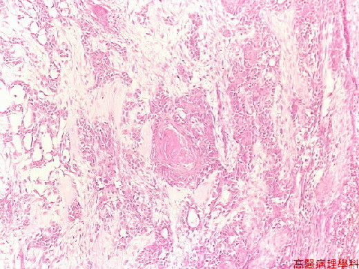
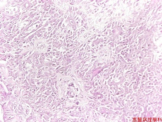
Epithelial component
-
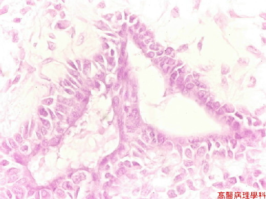
Glandular (ductal) with foci of squamous metaplasia.
-
Inner ductal lining cells from flatten to columnar.
-
Outer myoepithelial cells from cuboidal, flattened, clear spindle shaped or hyaline, sometimes proliferated to form thick, ill-defined sheaths around ducts, or become swollen & hydropic to cartilage-like cells (chondroid change).
-
Lumen may contain clear to eosinophilic fluid.
-
-
Stroma:Nonspecific fibromyxoid appearance with abundant elastic tissue (myxoid change), with clear-cut cartilaginous, myxochondroid, or calcification.
D. Others:
-
Most common neoplasm of the salivary glands especially parotid gland, with slow growth.
-
Proliferation of myoepithelial cells with pleomorphic appearance.
-
Some mimic epithelial component while others mimic stroma component (cartilage).
E. Reference:
Robbins Pathologic Basis of Disease, 6th ed. P.770-771.
|
|
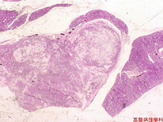
【 Fig. 36-1 (2X)】A relatively well defined tumor seen in this field.
|
|
【 Fig. 36-2 (1X)】A relatively well defined tumor seen in this field.
|
|
【 Fig. 36-3 (4X)】Biphasic appearance resulting from the admixture or epithelial & stroma.
|
|
【 Fig. 36-4 (10X)】Biphasic appearance resulting from the admixture or epithelial & stroma.
|
|
【 Fig. 36-5 (20X)】Epithelial component.
|
|
【 Fig. 36-6 (20X)】Epithelial component.
|
|
【 Fig. 36-7 (20X)】Epithelial component.
|
|
【 Fig. 36-8 (40X)】Glandular (ductal) component.
