《Slide 115.》Actinomycosis, Liver
A. Brief Descriptions:
-
Anaerobic non-spore forming bacteria, gram positive bacilli.
-
Normal commensal organism in the gastrointestinal tract.
-
Sulfar granules : yellowish granules found grossly.
B. Gross Findings:
-
Abscess contain sulfur granules.
C. Micro Findings:
-
Severely destructed liver with abscess formation.
-
In the abscess, sulfur granule with aggregation of Actinomycetic filaments.
D. Others:
-
Compare 《Slide 42.》 Actinomycosis of tongue.
E. Reference:
-
Robbins Pathologic Basis of Disease, 6th ed. P.369~370.
|
|
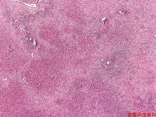
【 Fig. 115-1 (LP)】Focal destruction of hepatic parenchymal architecture.
|
|
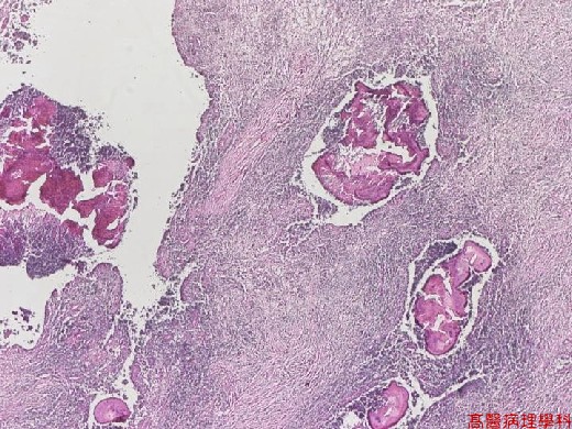
【 Fig. 115-2 (LP)】In abscess area, several dense basophilic sulfur granules are seen.
|
|
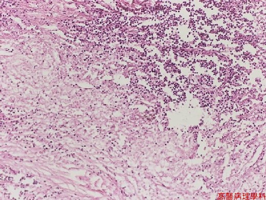
【 Fig. 115-3 (LP)】Foci of abscess formation showing necrotic debris accumulation with dense neutrophilic infiltration.
|
|
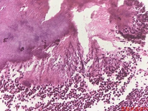
【 Fig. 115-4 (HP)】Radial arrangement of Actinomyceles with lymphocytic infiltrate at periphery.
![]()
![]()
![]()